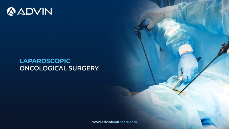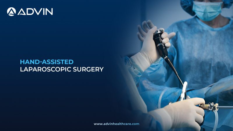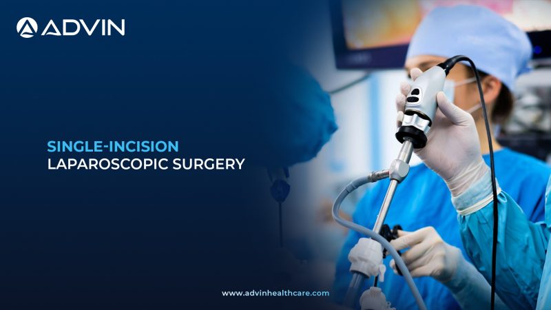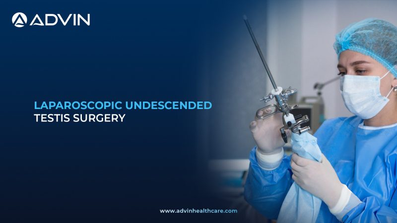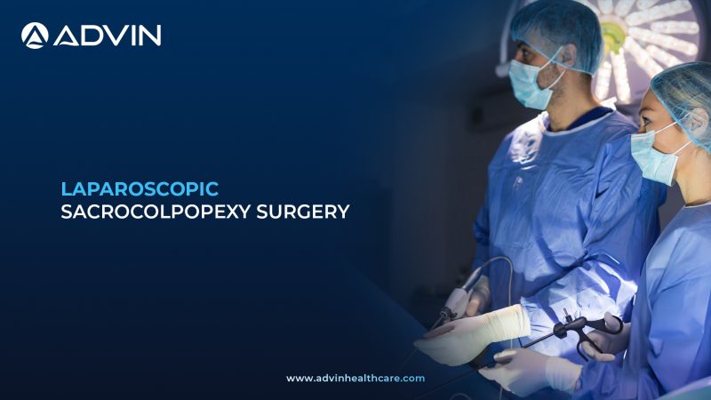
Overview of Laparoscopic Sacrocolpopexy Procedure
Laparoscopic sacrocolpopexy is a minimally invasive surgery used to treat pelvic organ prolapse. It restores the normal position of the vagina or uterus using mesh support. This procedure improves pelvic stability and function.
Minimally Invasive Laparoscopic Technique for Pelvic Organ Prolapse Repair
Laparoscopic sacrocolpopexy is performed under general anesthesia to ensure patient comfort and safety. Small abdominal incisions are made to insert a laparoscopic camera and surgical instruments. The vaginal vault or uterus is carefully exposed and prepared for suspension. A surgical mesh is attached to the vaginal wall and then fixed to the sacrum for strong support. Sutures or fixation devices are used to secure the mesh in proper position. This technique restores pelvic anatomy while preserving surrounding organs.
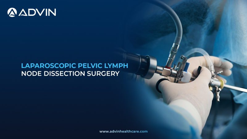
Clinical Indications for Laparoscopic Sacrocolpopexy
- To treat vaginal vault prolapse after hysterectomy
- To correct uterine prolapse in selected patients
- To relieve pelvic pressure and discomfort
- To improve bladder and bowel function affected by prolapse
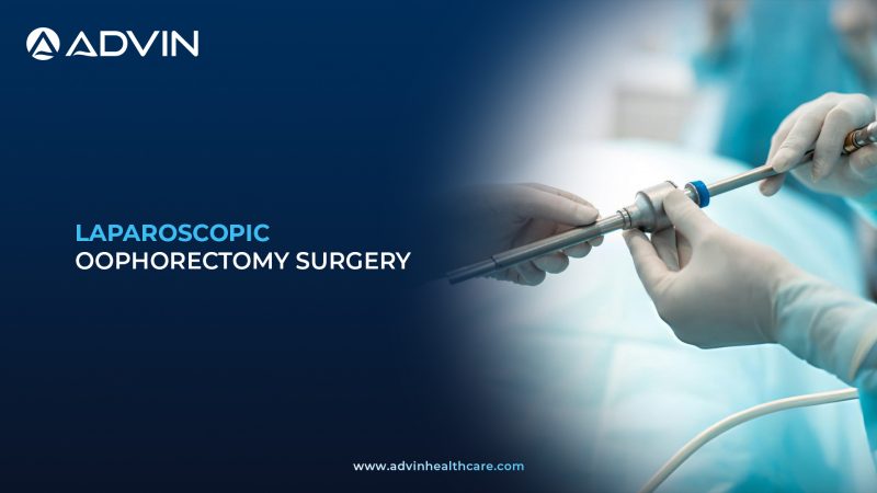
Laparoscopic Instruments, Meshes, and Accessories for Sacrocolpopexy
- Laparoscopic Camera System
- Laparoscopic Trocars
- Laparoscopic Graspers
- Laparoscopic Scissors
- Surgical Mesh
- Mesh Fixation Device or Sutures
- Laparoscopic Needle Holder
- Energy Sealing Device
- Suction Irrigation System
- Surgical Drapes
Leading Global Markets for Laparoscopic Sacrocolpopexy
- United States
- Germany
- Japan
- United Kingdom
- India
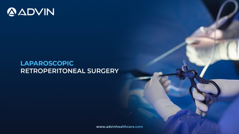
Clinical and Commercial Advantages of Laparoscopic Sacrocolpopexy
- Restores normal pelvic organ position
- Minimally invasive with smaller incisions
- Durable long-term support with low recurrence
- Improves urinary and bowel symptoms
- Faster recovery compared to open surgery
Advin Health Care is a globally leading manufacturer of laparoscopic sacrocolpopexy related products, providing reliable and high-quality solutions for advanced minimally invasive pelvic reconstructive procedures.
Get Connected:
+91-70717 27261 | urology@advinhealthcare.com | www.advinhealthcare.com