Anterior Cruciate Ligament Reconstruction

ACL reconstruction is surgery to replace a torn anterior cruciate (KROO-she-ate) ligament (ACL) — a major ligament in your knee. ACL injuries most commonly occur during sports that involve sudden stops and changes in direction — such as soccer, football, basketball and volleyball.

Ligaments are strong bands of tissue that attach one bone to another bone. During ACL reconstruction, the torn ligament is removed and replaced with a band of tissue that usually connects muscle to bone (tendon). The graft tendon is taken from another part of your knee or from a deceased donor.

ACL reconstruction is an outpatient surgery that’s performed by a doctor who specializes in surgical procedures of the bones and joints (orthopedic surgeon).

Why it’s done
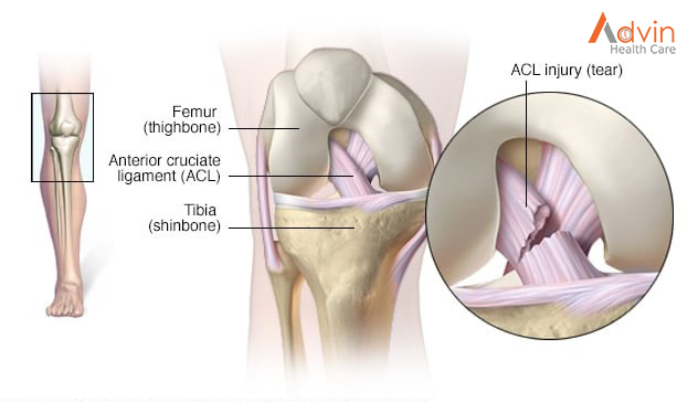

The ACL — one of two ligaments that crosses the middle of the knee — connects your thighbone to your shinbone and helps stabilize your knee joint.

Most ACL injuries happen during sports and fitness activities that can put stress on the knee:

  • Suddenly slowing down and changing direction (cutting)
  • Pivoting with your foot firmly planted
  • Landing from a jump incorrectly
  • Stopping suddenly
  • Receiving a direct blow to the knee

A course of physical therapy may successfully treat an ACL injury for people who are relatively inactive, engage in moderate exercise and recreational activities, or play sports that put less stress on the knees.

ACL reconstruction is generally recommended if:

  • You’re an athlete and want to continue in your sport, especially if the sport involves jumping, cutting or pivoting
  • More than one ligament is injured
  • You have a torn meniscus that requires repair
  • The injury is causing your knee to buckle during everyday activities
  • You’re young (though other factors, such as activity level and knee instability, are more important than age)

What Is ACL Surgery?
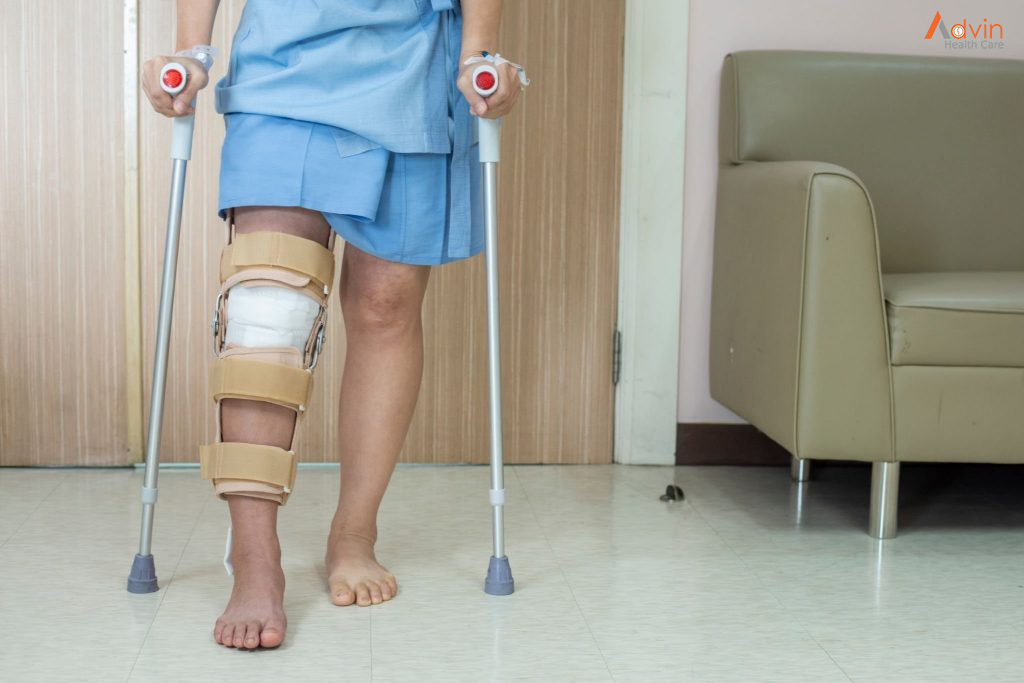

ACL surgery is a procedure that doctors use to replace a torn ligament in your knee.

The ACL (anterior cruciate ligament) is a band of tissue inside your knee. It gets damaged when it stretches or tears. ACL injuries are common among people who play sports because they make movements that can put a lot of stress on the knee, like:

  • Changing direction quickly (cutting)
  • Stopping suddenly
  • Planting your foot and pivoting
  • Landing wrong after a jump

When your ACL is healthy, it helps to hold together the bones of your knee. It also helps to keep your knee stable. If it gets damaged, you may have trouble putting pressure on your knee, walking, or playing sports.

If you strain or slightly tear your ACL, it may heal over time with your doctor’s help and physical therapy. But if it’s completely torn, you may need to have it replaced — especially if you’re young and active or an athlete who wants to keep playing sports. If you’re older or less active, your doctor might recommend treatments that don’t require surgery.

Types of ACL Surgery


When your doctor removes your torn ACL, they put a tendon in its place. (Tendons connect muscle to bone.) The goal is to get your knee stable again and give it the full range of motion it had before you got hurt.

When the tendon is put into your knee, it’s known as a graft. Three types of grafts can be used with ACL surgery:

  • Autograft. Your doctor uses a tendon from somewhere else in your body (like your other knee, hamstring, or thigh).
  • Allograft. This type of graft uses tissue from someone else (a deceased donor).
  • Synthetic graft. This is when artificial materials replace the tendon. Silver fibers and silk were among the first ones used (in the early part of the 20th century). More advanced options are available now, like carbon fiber and Teflon, but researchers are still working to find the best material for ACL replacement.

ACL Surgery Procedure


Doctors typically use arthroscopic surgery on your ACL. This means they insert tiny tools and a camera through small cuts around your knee. This method causes less scarring of the skin than open-knee surgery does.

The procedure takes about an hour. You may have general anesthesia, which puts you to sleep through the surgery, or you may have regional anesthesia, when your doctor puts medicine in your back so you won’t feel anything in your legs for a few hours. If you have regional anesthesia, you probably will also get medicine that helps you relax during the procedure.

The first step is to place the graft at the right spot. Then, your doctor will drill two holes, called “tunnels.” They’ll put one in the bone above your knee and another in the bone below it. They’ll place screws in the tunnels to hold the graft in place. It serves as a sort of bridge that a new ligament will grow on as you heal. It can take months for a new ACL to grow in fully.

After surgery, most people are able to go home the same day. Your doctor will have you stay off your leg, rest your knee, and wear a brace to protect the joint.

Doctors are also involved in research to see if a new type of ACL surgery is better than the standard care. It’s called bridge-enhanced ACL repair (BEAR).

Advin Products


ACL PCL Instrument Set

The Facts About Liver Transplant

A liver transplant is a surgery that removes a liver that no longer functions properly (liver failure) and replaces it with a healthy liver from a deceased donor or a portion of a healthy liver from a living donor.

Your liver is your largest internal organ and performs several critical functions, including:

  • Processing nutrients, medications and hormones
  • Producing bile, which helps the body absorb fats, cholesterol and fat-soluble vitamins
  • Making proteins that help the blood clot
  • Removing bacteria and toxins from the blood
  • Preventing infection and regulating immune responses

Liver transplant is usually reserved as a treatment option for people who have significant complications due to end-stage chronic liver disease. Liver transplant may also be a treatment option in rare cases of sudden failure of a previously healthy liver.

The number of people waiting for a liver transplant greatly exceeds the number of available deceased-donor livers.

Receiving a portion of a liver from a living donor is an alternative to waiting for a deceased-donor liver to become available. Living-donor liver transplant is possible because the human liver regenerates and returns to its normal size shortly after surgical removal of part of the organ.

What is a liver transplant?


Liver transplant is a surgery performed to replace one’s damaged liver with a whole or partial healthy liver from another person. It is done to save a person’s life. Healthy liver constitutes a healthy and happy life.

The liver is the largest internal organ and performs hundreds of different functions for the body, which include fighting infections, detoxifying all the chemicals, and manufacturing protein and minerals. The liver holds approximately 10% of the blood in your body and it is the only organ to regenerate or regrow by itself. So if someone donates half of their liver to the person who needs a transplant, his/her liver will return to its normal shape and size in almost 2 weeks. Therefore, a liver transplant gives a new life and a new ray of sunshine to a person suffering from any liver disease.

Why liver transplants are done?


A liver transplant is done when someone is suffering from liver failure or liver cancer. Liver failure can either occur quickly or after a long period of time. Acute liver failure is a condition where failure occurs quickly in a matter of weeks. Acute liver failure may need a transplant but it is often used to treat chronic liver failure which starts slowly or may take months and years to get noticed.

One of the common causes of chronic liver failure is a condition called cirrhosis. Cirrhosis is the last stage of liver disease where a healthy liver gets replaced with scarred tissue (fibrosis) caused mainly due to certain conditions like hepatitis and extreme alcoholism and it permanently damages the liver. Hence, a patient will be needed an immediate liver transplant to cure cirrhosis. Other causes of cirrhosis are:

  • Nonalcoholic fatty liver disease
  • Hemochromatosis (Iron buildup in the body)
  • Wilson’s disease (accumulation of copper in the liver)
  • Autoimmune hepatitis
  • Biliary Artesia ( poorly formed bile ducts)
  • Genetic digestive disorder
  • Cystic fibrosis

Who requires a Liver Transplant?


If you are facing any liver-related disease or if your liver stops working then you need a liver transplant. The transplant is needed only when the condition is serious and life-threatening and you reach the end-stage of the disease. You might require a liver transplant which you are suffering from cirrhosis. The causes are mentioned above. Also if you have liver cancer, the doctor can suggest a transplant.

Possible risks and complications of a liver transplant


Any surgery or transplant comes with possible risks and complications that might arise. It is not necessary that all transplant comes with risk. In the case of a liver transplant, there are a few below-enlisted risks and complications during or after a transplant.

  • Rejection of the donated liver, Sometimes after a transplant, the body rejects the donated liver or starts attacking the liver.
  • Blood clots
  • Bleeding
  • Damage to the bile ducts, During a liver transplant, one of your bile ducts can suffer blockage or leak.
  • Failure of the donated liver, There is a chance that the new liver stops responding to the body or does not work properly.
  • Infection, After a liver transplant surgery, many face infection as it takes time for a body to accept the new liver.

Healthy liver tips


A healthy liver is equal to a healthy and happy you. It becomes extremely important to keep your body and its internal organs healthy which helps in keeping all the diseases at bay. Our health should always be our first priority and especially the liver which handles all-important functions of the body. Here are few tips to keep your liver healthy. These tips are known to all but we fail to practice or follow.

Maintain a balanced diet

Always eat a balanced diet with all necessary quantities of nutrients like vitamins, minerals, carbohydrates, and fats. Have an equal amount of fibers that you obtain from fruits, vegetables, seeds, etc. Leafy Vegetables also help to keep your liver healthy. Avoid raw or processed foods, high saturated fats, refined carbohydrates like wheat, all-purpose flour, and also cut down on sugar intake. Also have meats, dairy products, and good fats. Most important than all is water. Hydration is essential for any bodily activity to work smoothly. Have almost 10-12 glasses of water per day.

Exercise regularly

Exercise is essential to maintain a healthy life. Practice yoga or exercise regularly in the morning and evening to cut down on liver fat or burn any other fats in the body.

Maintain a healthy body weight

One of the symptoms to look for liver disease is an increase in body weight. So if your overweight or obese, try to lose weight as it can constitute more complicated liver disease.

Avoid any junk or toxic foods

Avoid junk foods that contain a large amount of gluten, sugar, and unsaturated fats. Junk or any toxic foods automatically deteriorates the overall health.

Limit your alcohol and other drinks intake

Alcohol is very dangerous to health. Intake of alcohol or any other beverages can anyway destroy your liver and kidney. Try to limit the intake and talk to the doctor about the amount of alcohol one can limit to keep the liver safe from diseases.

Cut down on smoking

Smoking not only affects respiratory organs but also the liver. Smoking cigarettes in high amounts is cancerous as it contains nicotine and tar which are harmful to our body. Try to stop smoking or limit the intake.

Limit the use of drugs

Drugs like marijuana, cocaine, heroin, inhalants are toxic to the body and can give rise to many liver complications.

Be aware of certain medicines

Always have the prescribed medicines given by the doctor consulted in the given time period and in a given quantity. Don’t consume any other traditional medicines without consulting the doctor as it can adverse effects.

Get vaccinated for hepatitis A and B

There are vaccines for Hepatitis A and Hepatitis B. These vaccines help to curb the disease and it becomes extremely important for all to get vaccinated.

Schedule a regular visit to a doctor.

Even if you are not facing any issues, having a regular checkup is necessary because there are few diseases that are asymptomatic and early diagnosis can help it cure in early stage.

Thyroidectomy Surgery

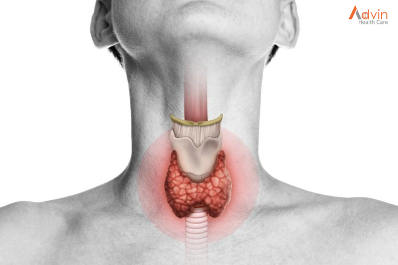
Thyroidectomy is the surgical removal of all or part of your thyroid gland. Your thyroid is a butterfly-shaped gland located in the front of your neck. It makes hormones that control every part of your metabolism, from your heart rate to how quickly you burn calories.

Health care providers perform thyroidectomy to treat thyroid disorders. These include cancer, noncancerous enlargement of the thyroid (goiter) and overactive thyroid (hyperthyroidism).

How much of your thyroid gland is removed during thyroidectomy depends on the reason for the surgery. If you need only part of your thyroid removed (partial thyroidectomy), your thyroid may work normally after surgery. If you need your entire thyroid removed (total thyroidectomy), you need daily treatment with thyroid hormone to replace your thyroid’s natural function.

What is a thyroidectomy?


A thyroidectomy is the surgical removal of all (total thyroidectomy) or part (partial thyroidectomy) of your thyroid gland — the butterfly-shaped organ in your neck.

Thyroidectomy is the main surgical treatment for thyroid cancer and is a treatment option for certain thyroid conditions, including:

  • Thyroid nodules: A thyroid nodule is a growth (lump) of thyroid cells in your thyroid gland. Thyroid nodules are usually benign (noncancerous), but they can be malignant (cancerous). Sometimes, thyroid nodules can produce excess thyroid hormone, which causes certain symptoms.
  • Goiter: Goiter is an enlarged thyroid gland with or without thyroid nodules. If it grows large enough, it can put pressure on your trachea or food pipe (esophagus) and make it more difficult to breathe and swallow.
  • Hyperthyroidism: Hyperthyroidism (overactive thyroid) is a condition in which your thyroid creates and releases more hormones than you need. It has several causes, and surgery is one of the treatment options for the condition.

Why it’s done


Your doctor may recommend thyroidectomy if you have conditions such as:

  • Thyroid cancer. Cancer is the most common reason for thyroidectomy. If you have thyroid cancer, removing most or all of your thyroid will likely be a treatment option.
  • Noncancerous enlargement of the thyroid (goiter). Removing all or part of your thyroid gland may be an option for a large goiter. A large goiter may be uncomfortable or make it hard to breathe or swallow. A goiter may also be removed if it’s causing your thyroid to be overactive.
  • Overactive thyroid (hyperthyroidism). In hyperthyroidism, your thyroid gland produces too much of the hormone thyroxine. Thyroidectomy may be an option if you have problems with anti-thyroid drugs, or if you don’t want radioactive iodine therapy. These are two other common treatments for hyperthyroidism.
  • Suspicious thyroid nodules. Some thyroid nodules can’t be identified as cancerous or noncancerous after testing a sample from a needle biopsy. If your nodules are at increased risk of being cancerous, you may be a candidate for thyroidectomy.

Types of Thyroid Operations


In general, there are three types of thyroid resections:

Total thyroidectomy: removal of the entire thyroid

A total thyroidectomy may be done for a variety of diseases including thyroid cancer, Graves’ disease (See Hyperthyroidism »), multinodular goiter, and substernal goiter, among others. In certain cases, the surgeon may choose to perform a near-total thyroidectomy in which a small piece of thyroid tissue is left behind usually in the area of the parathyroid glands and recurrent laryngeal nerve in order to avoid damaging these structures. After a total thyroidectomy, patients will need to take thyroid hormone replacement pills (one pill a day for the rest of their lives).

Thyroid lobectomy (aka hemithyroidectomy): removal of half of the thyroid

A thyroid lobectomy may be done for a variety of diseases including indeterminate lesions on fine needle biopsy (See Thyroid Nodules Diagnosis and Treatment »), a toxic nodule (See Hyperthyroidism »), substernal goiter, and an enlarging thyroid nodule, among others. In cases of indeterminate lesions, some surgeons refer to a thyroid lobectomy as a diagnostic lobectomy because the main purpose of the operation is to make a diagnosis – cancer or benign thyroid disease. The final pathology is ready approximately 1 week after the operation. Approximately 70% of patients who have half of a normal thyroid gland left in place will not require thyroid hormone replacement pills. This percent decreases in older women, patients with a personal or family history of Hashimoto’s thyroiditis or hypothyroidism, and patients with a family history of autoimmune disease.

Completion thyroidectomy: removal of any remaining thyroid tissue.

A completion thyroidectomy is usually done after a thyroid lobectomy reveals cancer in the first half of the thyroid but may also be done for multinodular goiter or hyperthyroidism. After a completion thyroidectomy, patients will need to take thyroid hormone replacement pills (one pill a day for the rest of their lives).

The decision as to which thyroid operation to perform depends on a number of factors including the type of disease and the patient’s preferences. It is critical to work with a team of thyroid specialists to choose the right operation for each individual patient.

Advin Thyroidectomy Instrument set


Ankle Fracture Surgery

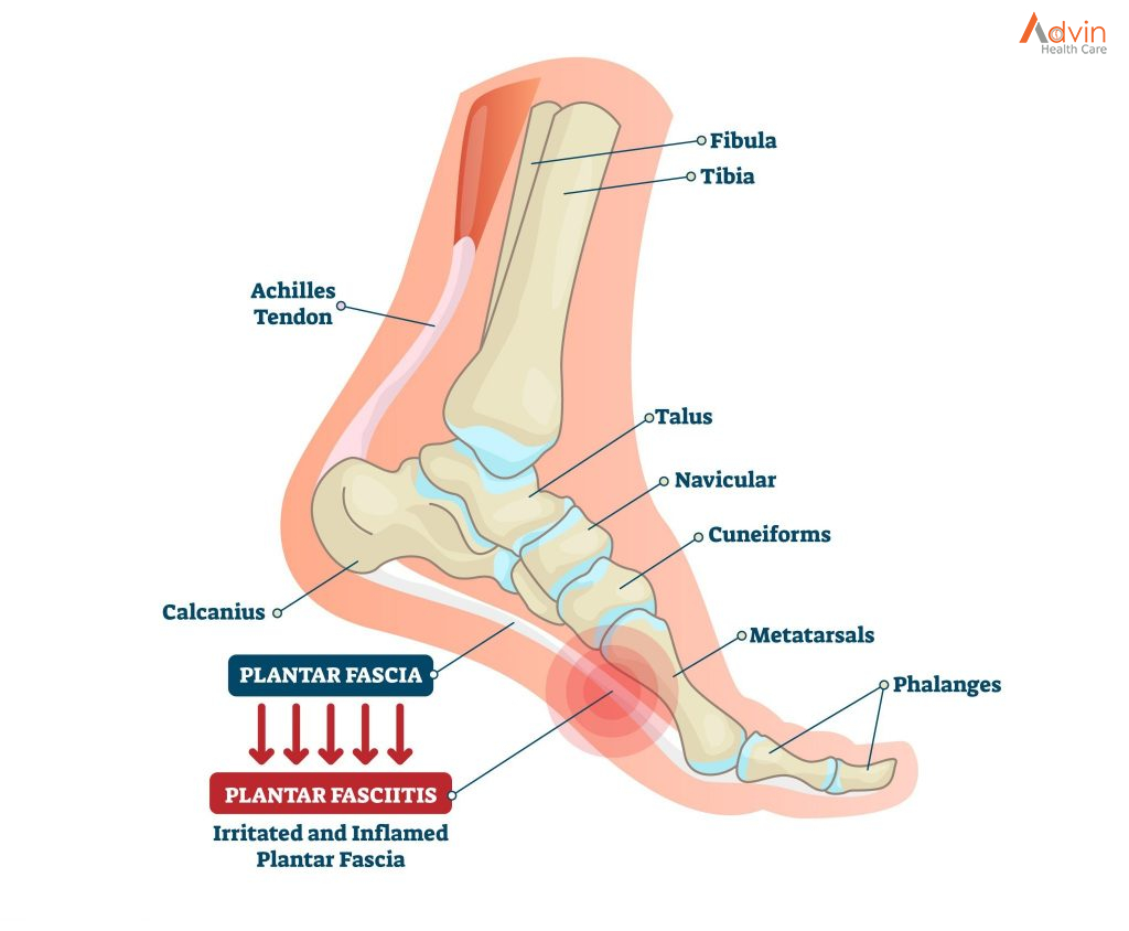
The ankle is made up of three bones:

the tibia (shin bone), which forms the inside, front, and back of the ankle

the fibula, which forms the outside of the ankle

the talus, a small bone that sits between the tibia and fibula and the heel bone

Parts of the ankle

The ends of these bones are called malleoli. The tibia has a medial (inside) malleoli and a posterior malleolus. The fibula forms the lateral (outside) malleoli.

A broken ankle (ankle fracture) occurs when the malleoli are broken. These fractures are very common. Ankle fractures happen with twisting of the ankle, falls, car accidents, or other injury. One, two, or all three malleoli can be broken. Ankle fractures can be displaced (out of place) or non-displaced. Symptoms of an ankle fracture include pain especially with weight bearing, swelling, bruising, and problems with ankle motion. X-rays help determine if treatment from a foot and ankle orthopaedic surgeon is needed.

The main goal of ankle fracture surgery is to put the ankle joint back in place and to stabilize the bones to heal. Getting the ankle joint back in place helps to decrease the risk of developing arthritis of the ankle. Stabilizing the ankle with plates and screws may allow earlier motion.

Ankle Fracture Symptoms


If you’re experiencing any of the following symptoms, it is very important that you get checked by your doctor to determine if you have a fracture:

  • Throbbing pain
  • Increased pain during activity and decreased pain during rest
  • Bruising
  • Inflammation, redness and tenderness
  • A deformity of the bone in the foot and/or ankle
  • Difficulty walking
  • Difficulty bearing weight

How to Diagnose Ankle Fractures


To diagnose a foot and ankle fracture, you should be seen by an orthopaedic specialist to assess the severity of the injury and determine a plan for treatment. Imaging tests will likely be ordered, which may include:

  • Musculoskeletal ultrasound
  • MRI
  • CT scans
  • Weight-bearing CT scan

There are several types of fractures:

  • Comminated fracture: the bone is broken into more than two pieces
  • Extra-articular fracture: the break does not extend into the joint
  • Intra-articular fracture: the break extends into the joint
  • Open fracture: the fractured bone breaks the skin

Ankle Fracture Treatment Options

In some cases, if the fracture is mild and non-displaced (meaning the bone hasn’t shifted out of place), non-surgical treatment may be able to heal it. To treat foot fractures and ankle fractures without surgery, we typically follow the RICE protocol:

  • Rest: Rest is key. Staying off your injury will help you heal faster. You will likely wear a cast to help keep the foot and ankle immobilized.
  • Ice: Ice the area for 20 minutes at a time to help with swelling and inflammation. Continue icing every 40 minutes.
  • Compression: Wrapping the injury helps control swelling.
  • Elevation: Keep your foot and ankle raised slightly above the level of your heart to help reduce inflammation.

Are there different types of ankle fractures?


There are several types of ankle fractures affecting different parts of your ankle. Ankles are complicated. They’re made up of three bones and four ligaments, each doing a different job to keep your ankle in good working order. Here are the different types of ankle fractures:

  • Lateral malleolus fractures: This injury can happen when you break the bony knob on the outside of your ankle. This is the most common type of ankle fracture.
  • Medial malleolus fractures: This fracture happens when you break the bony knob on the inside of your ankle.
  • Bimalleolar ankle fracture: This fracture happens when you break both bony knobs on your ankle. This is the second most common type of ankle fracture.
  • Bimalleolar equivalent fracture: This fracture happens when you break both bony knobs on the outside of your ankle and you damage ligaments inside your ankle.
  • Posterior malleolus fracture: There’s a bony section on the back of your tibia. This is your posterior malleolus.
  • Trimalleolar fracture: In this case, all three parts of your ankle are broken.
  • Pilon fracture: Your tibia ends in a section called the roof of your ankle. When you break this section, it’s called a Pilon fracture.
  • Maisonneuve fracture: This fracture happens when you sprain your ankle and break the upper part of your fibula, near your knee.
  • Syndesmotic injury: The syndesmosis joint is located between your fibula and tibia (shinbone) and anchored by ligaments. A syndesmotic injury happens when you have at least one fracture in your tibia or fibula and you sprain ligaments in your syndesmotic joint.

Ankle Fracture Surgery


If you have a more serious fracture, surgery is usually necessary to repair the fracture as well as any ligaments, tendons and muscles that have been damaged. Our orthopaedic surgeons are experts in fracture repair of the foot and ankle. We have the expertise to treat complex cases, including broken bones that haven’t healed properly (called non-union) and other types of traumatic fractures.

For complex wounds of the foot, we have a unique program called the Orthoplastic Limb Salvage Center, where a specialized team of orthopaedic surgeons and plastic surgeons work together to determine a plan for reconstructive surgery and operate together. Orthopaedic foot and ankle surgeons have expertise in treating all types of bone fractures and performing bone-grafting procedures, while plastic surgeons have specialized training and knowledge of soft tissue injuries that may accompany traumatic fractures.

The Penn Orthoplastic Limb Salvage Center is the only program of this kind in the U.S. that provides this level of integrated care and expertise in microsurgical and complex fracture treatment. All of the surgeons are accustomed to treating injuries that require multiple surgeries at the same time, including re-plantation of limbs and toes and specialized microvascular procedures to repair bones and soft tissues. We consistently combine highly advanced surgical specialties that ensure the best possible outcome for more serious fractures and wounds of the foot and ankle and prevent limb amputation for those at risk.

Some of the surgical treatments for foot and ankle fractures that we often perform are fixation surgery, non-union surgical repair and reduction surgery.

Ankle Fracture Fixation Surgery

If the fracture in your foot or ankle is displaced, or has caused misalignment of the bones, your orthopaedic surgeon will need to put the bones back into the correct position using specialized hardware such as plates, screws or pins. If necessary, we will perform bone grafting and may need to reconstruct soft tissues such as ligaments and tendons.

Non-Union Ankle Fracture Surgery

A non-union is a broken bone that did not heal properly. Treatment may entail surgery to remove an infection if present, to better stabilize the fracture, or to stimulate bone growth with a bone graft.

Our surgeons perform complex non-union surgical repair with vascularized bone grafting. Vascularized bone grafts allow living bone tissue to be transplanted to replace bone tissue that is damaged.

We are one of the few medical centres in the nation who perform vascularized bone grafting.

Ankle Reduction Surgery

If the fracture in your foot or ankle is displaced, or has caused misalignment of the bones, your orthopaedic surgeon will need to put the bones back into the correct position using specialized hardware such as plates, screws or pins. If necessary, we will perform bone grafting and may need to reconstruct soft tissues such as ligaments and tendons.

Advin Ankle Instrument Set


Open Heart Surgery

Open heart surgery is an umbrella term for various procedures that involve opening up a person’s ribcage through a large chest incision in order to expose their heart. This is a major operation that may be performed for a number of reasons including bypassing a blocked heart artery, repairing a diseased heart valve, or transplanting a healthy heart.

While the surgery offers many benefits and is often life-changing, recovery is usually gradual and challenging. After surgery, patients must be firmly committed to their follow-up care and heart-healthy lifestyle habits.

What Is Open Heart Surgery?


Open heart surgery is an inpatient operation performed by a cardiothoracic surgeon or heart transplant surgeon in a hospital under general anesthesia. This type of surgery may be scheduled or performed emergently, depending on the patient’s medical circumstances.

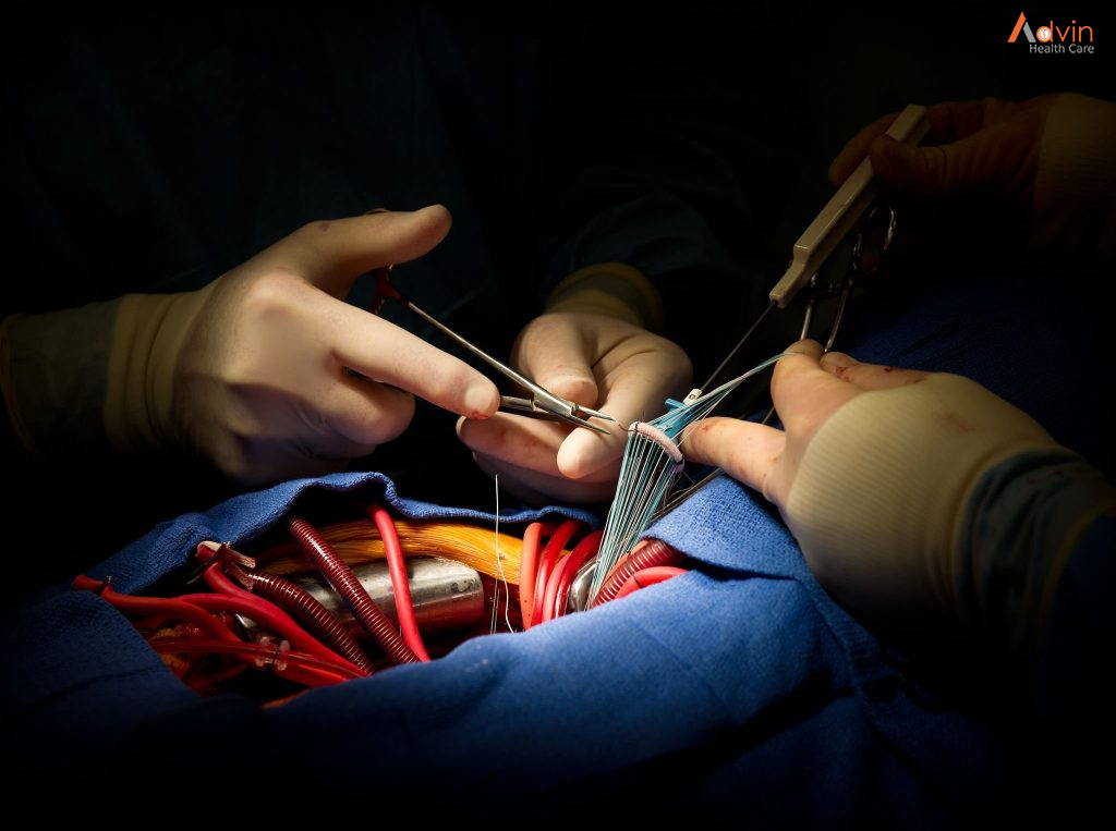
During open heart surgery, the surgeon makes a large incision in the middle of the patient’s chest. The breastbone (which is connected to the ribcage) is then cut in half lengthwise and spread apart in order to expose the heart within the chest cavity.

Once the heart is exposed, the patient is connected to a heart-lung bypass machine. This machine takes over the heart’s function, pumping oxygen-rich blood throughout the body. As a result, the surgeon is able to operate on a “still” heart (i.e., one that is not beating and has no blood flowing through it).

Open heart surgery may be performed for many different reasons. Most commonly, it is used to bypass a diseased heart (coronary) artery—what’s called a coronary artery bypass graft (CABG).

Various Surgical Techniques


Even though open heart surgery is an invasive technique, it’s still widely used as it allows the surgeon to directly visualize the heart and its surrounding blood vessel supply.

That said, over the years, various minimally invasive approaches have emerged. These alter what most expect of open heart surgery in some key ways and may or may not be viable options, depending on the case.

For example, with the minimally invasive direct coronary artery bypass (MIDCABG) approach, the surgeon makes several small incisions on the left side of the chest in order to reach and operate on a patient’s coronary arteries.2

With this approach, most patients are not placed on a heart-lung machine.3 This means that the flow of blood through the body is maintained by the heart during the procedure; this is called “off-pump” surgery.

There are also minimally invasive valve repair or replacement techniques. With minimally invasive mitral valve surgery, for instance, a small incision is made on the right side of the chest.4

A specialized instrument is then inserted through the small opening and used to repair the valve.

Purpose of Open Heart Surgery


Open heart surgery is used to treat a number of different heart conditions.

Coronary artery disease (CAD), the most common indication, occurs when fatty clumps (plaques) clog the arteries that supply blood to the heart muscle. This results in reduced blood flow to the heart. If the blockage is significant, angina, trouble breathing, and, in some cases, a heart attack may occur.

Open heart surgery may also be used to:

  • Treat end-stage heart failure
  • Treat refractory heart arrhythmias, including atrial fibrillation (called the Maze heart procedure)
  • Repair diseased/damaged heart valves
  • Repair congenital heart defects
  • Treat cardiomyopathy (enlarged heart)
  • Implant a medical device, like a left ventricular assistive device (LVAD)
  • Transplant a heart

The benefits of open-heart surgery are often immense and can include:

  • Alleviating or reducing symptoms like chest pain or trouble breathing
  • Decreasing the risk of cardiovascular conditions like stroke or heart attack
  • Improving survival and quality of life14

Several different pre-operative tests are performed before a patient undergoes open heart surgery.

Examples of these tests include:

  • Blood tests, such as complete blood count (CBC) and a coagulation panel
  • Electrocardiogram (ECG or EKG)
  • Echocardiogram
  • Cardiac stress test
  • Cardiac catheterization
  • Pulmonary function tests
  • Psychological and social evaluation (for a heart transplant)

How to Prepare


Once an open heart surgery is scheduled, your surgeon will give you specific instructions to follow.

These may include:

  • Avoid chewing, eating, or drinking anything (including water) after 10 p.m. on the eve of your surgery.
  • Avoid caffeine and alcohol up to 48 hours before surgery.16
  • Shower with a special antibacterial soap starting two to four days prior to surgery.
  • Stop smoking and/or tobacco use as soon as possible.
  • Cease or continue certain medications prior to surgery.
  • Undergo several vaccinations (applicable only to patients undergoing a heart transplant).

What to Expect on the Day of Surgery


Prepping you for surgery will involve several steps, including checking your vitals, placing an IV in your hand, arm, or neck to deliver fluids and medications during surgery, and placing an arterial line (a thin catheter that goes in an artery in your wrist) to monitor your blood pressure.

Once you are in the operating room, an anesthesiologist will give you medications to put you to sleep and a breathing (endotracheal) tube will be inserted. This tube is connected to a ventilator to assist with breathing during surgery.

A Foley catheter will also be placed at this time to drain urine. In select cases, the surgeon may place a thin tube called a Swan-Ganz catheter in a vein in your neck. This catheter measures pressures in and around the heart and is used for monitoring purposes during and right after surgery.

The precise steps of your open heart surgery depend on what exactly is being done (valve repair, heart transplant, coronary artery bypass, etc.) and what technique is being used.

That said, here is a general breakdown of a traditional open heart surgery:

  • Access: The skin over your chest will be cleaned, the surgeon will make an 8-inch incision down the center of the chest wall, and then cut your breastbone in half lengthwise. The breastbone will be separated from your ribs and spread apart to expose the heart.
  • Operating on the heart: You will be placed on a heart-lung bypass machine. Then, depending on the operation, various steps will be performed. For example, one or more blocked coronary arteries may be bypassed during a CABG. A diseased valve may be removed and replaced with an artificial valve during a valve replacement.
  • Completion: The surgeon will remove you from the bypass machine to allow blood flowing through it to re-enter your heart. Temporary pacemaker wires placed during surgery (if applicable) will be attached to the device outside your body. The breastbone will then be sewn back together with small wires. Chest tubes may be placed to drain blood and other fluids around the heart.17 Muscles and skin are then closed with sutures. A sterile bandage will be applied over the large incision site.

Lumpectomy Surgery

Lumpectomy (lum-PEK-tuh-me) is surgery to remove cancer or other abnormal tissue from your breast.

During a lumpectomy procedure, the surgeon removes the cancer or other abnormal tissue and a small amount of the healthy tissue that surrounds it. This ensures that all of the abnormal tissue is removed.

Lumpectomy is also called breast-conserving surgery or wide local excision because only a portion of the breast is removed. In contrast, during a mastectomy, all of the breast tissue is removed. Doctors may also refer to lumpectomy as an excisional biopsy or quadrantectomy.

Lumpectomy is a treatment option for early-stage breast cancer. Sometimes lumpectomy is used to rule out a cancer diagnosis. When a lumpectomy surgery is performed to remove cancer, it usually is followed by radiation therapy to the breast to reduce the chances of cancer returning.

Why it’s done


The goal of lumpectomy is to remove cancer or other abnormal tissue while maintaining the appearance of your breast. Studies indicate that lumpectomy followed by radiation therapy is as effective in preventing a recurrence of breast cancer as removal of the entire breast (mastectomy) for early-stage breast cancer.

Your doctor may recommend lumpectomy if a biopsy has shown that you have cancer and that the cancer is believed to be small and early stage. Lumpectomy may also be used to remove certain noncancerous or precancerous breast abnormalities.

Your doctor may not recommend lumpectomy for breast cancer if you:

  • Have a history of scleroderma, a group of diseases that harden skin and other tissues and make healing after lumpectomy difficult
  • Have a history of systemic lupus erythematosus, a chronic inflammatory disease that can worsen if you undergo radiation treatments
  • Have two or more tumors in different quadrants of your breast that cannot be removed with a single incision, which could affect the appearance of your breast
  • Have previously had radiation treatment to the breast region, which would make further radiation treatments too risky
  • Have cancer that has spread throughout your breast and overlying skin, since lumpectomy would be unlikely to remove the cancer completely
  • Have a large tumor and small breasts, which may cause a poor cosmetic result
  • Don’t have access to radiation therapy

What should I expect before a lumpectomy?


The details of your case (such as a tumor’s size or location) can affect how your breast looks after surgery. Your surgeon will review your imaging with the radiologist and design a surgical procedure which will focus on removing the entire cancer area.

In some cases, due to the larger size of the tumor, the breast surgeon may enlist the assistance of a plastic/reconstructive surgeon to perform combined team approach surgery called an oncoplastic lumpectomy. In an oncoplastic operation, the cancer is a bit bigger than suitable for a traditional lumpectomy (removing more than 20% of the breast volume), so the planned surgery takes into account the larger piece of breast tissue needing to be removed and allows for better scar placement, reshaping and nipple position. Often oncoplastic surgery requires a surgery on the other, normal breast to reduce the volume as well to match the cancer breast size.

It’s important to talk with your healthcare provider before surgery about how your breast may change. Your provider can tell you what breast surgery options are available.
Your provider will go over procedure details before surgery to help you feel comfortable about your care. Ask your provider to explain anything you don’t understand or are unsure about.
It’s important for your health and safety that you follow all of your provider’s instructions, such as when to stop taking certain medications before surgery.

Locating the area to be removed

Your lumpectomy procedure begins with locating the area of your breast that contains the abnormality (localization procedure). To do this, a doctor who uses imaging tests to diagnose and treat diseases (radiologist) uses a mammogram or ultrasound to locate the tumor and insert a thin wire, needle or small radioactive seed. Your surgeon uses this as a guide to find the precise area that needs to be removed during surgery.

If you have a lump or mass in your breast that can be easily felt through the skin, the localization procedure may not be necessary because the surgeon can more easily find the abnormal area to be removed.

Preparing for lymph node removal

Your surgeon may recommend removing lymph nodes near your armpit to see whether cancer has spread beyond the breast. Surgery to remove a few lymph nodes (sentinel node biopsy) is often recommended for early-stage breast cancer. If cancer was found in a lymph node before surgery or if there’s a concern that the cancer has spread, your surgeon may recommend removing a number of lymph nodes near your armpit (axillary lymph node dissection).

Lymph node removal procedures include

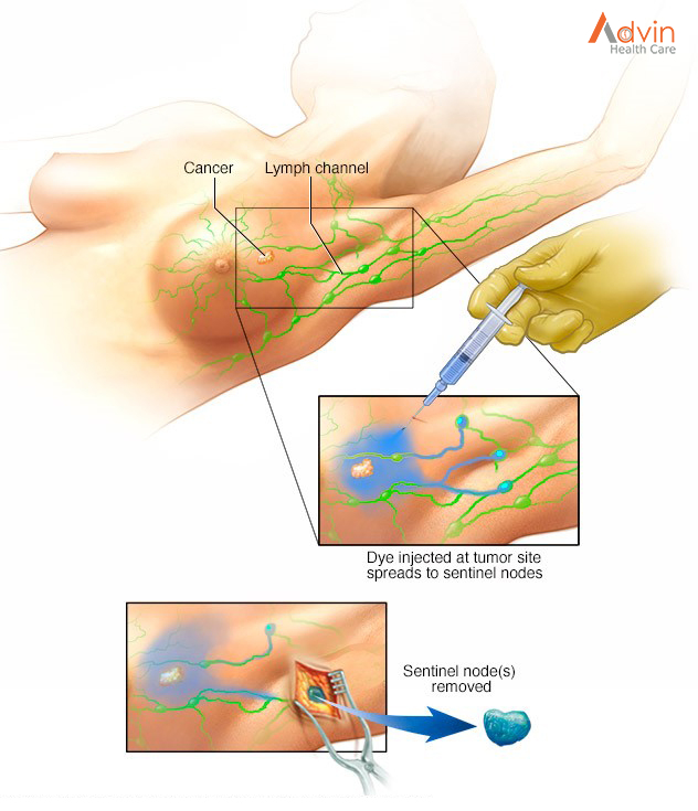
Sentinel node biopsy. During this procedure your surgeon removes only the first one or two nodes into which the cancer drains (sentinel nodes). These are then tested for cancer. Your doctor may recommend this procedure if there are no concerns about enlarged lymph nodes prior to your surgery.

Before your surgery, a radioactive substance or blue dye or both is injected into the area around the cancer or the skin above the cancer. The dye travels to the sentinel node or nodes, allowing your surgeon to identify and remove them.

If no cancer is present in the lymph nodes, no further lymph nodes need to be removed. If cancer is present, the surgeon will discuss options, such as receiving radiation to the armpit. If this is what you decide to do, you will not need to have more lymph nodes in the armpit removed.

Axillary lymph node dissection. During this procedure, the surgeon removes a number of lymph nodes from your armpit. Your surgeon may recommend this procedure if a lymph node biopsy done before surgery shows signs of cancer.

During the procedure


A lumpectomy is usually performed using general anesthesia, which will put you into a sleep-like state during the procedure.

Your surgeon will make an incision over the tumor or over the area that contains the wire or seed, remove the tumor and some surrounding tissue, and send it to the lab for analysis. He or she will do the same for the sentinel lymph node or nodes if you’re having a sentinel node biopsy or the axillary lymph nodes if you’re having an axillary lymph node dissection.

The surgeon will then close the incisions with attention to preserving the appearance of your breast, using stitches (sutures) that will either dissolve on their own or be removed later by your doctor. Your surgeon may also place thin adhesive strips or use glue on the incision to keep it closed until it heals.

After the procedure


After your surgery, you’ll be taken to a recovery room. During this time, your blood pressure, pulse and breathing will be monitored.

If you’ve had outpatient surgery — usually lumpectomy and sentinel node biopsy — you’ll be released when your condition is stable.

If you’ve had axillary lymph node dissection, you may need to stay in the hospital for a day or two if you’re experiencing pain or bleeding.

Expect to have:

  • A dressing (bandage) over the surgery site
  • Some pain, numbness and a pinching sensation in your underarm area
  • Written instructions about post-surgical care, including caring for the incision and dressing and recognizing signs of infection
  • Prescriptions for pain medication and possibly an antibiotic
  • Some restrictions of activity
  • A follow-up appointment with your doctor, usually seven to 14 days after surgery

Advin Lumpectomy Set


Bariatric Surgery

Bariatric surgery is a term that refers to surgical procedures that assist in weight loss by making changes to the digestive system. The term “bariatric surgery” can be used to describe a variety of procedures, such as gastric bypass and other weight loss surgeries.

Gastric bypass and other weight-loss surgeries — known collectively as bariatric surgery —involve making changes to your digestive system to help you lose weight. Bariatric surgery is done when diet and exercise haven’t worked or when you have serious health problems because of your weight. Some procedures limit how much you can eat. Other procedures work by reducing the body’s ability to absorb nutrients. Some procedures do both.

While bariatric surgery can offer many benefits, all forms of weight-loss surgery are major procedures that can pose serious risks and side effects. Also, you must make permanent healthy changes to your diet and get regular exercise to help ensure the long-term success of bariatric surgery.

Sleeve Gastrectomy


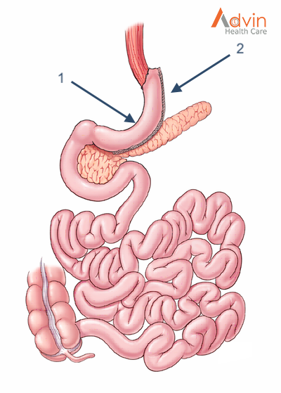
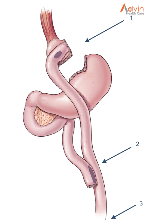
The Laparoscopic Sleeve Gastrectomy, often called the “sleeve”, is performed by removing approximately 80% of the stomach. The remaining stomach is the size and shape of a banana.

The Procedure
  1. The stomach is freed from organs around it.
  2. Surgical staplers are used to remove 80% of the stomach, making it much smaller.
How it Works

The new stomach holds less food and liquid helping reduce the amount of food (and calories) that are consumed. By removing the portion of the stomach that produces most of the “hunger hormone”, the surgery has an effect on the metabolism. It decreases hunger, increases fullness, and allows the body to reach and maintain a healthy weight as well as blood sugar control. The simple nature of the operation makes it very safe without the potential complications from surgery on the small intestine.

Advantages
  1. Technically simple and shorter surgery time
  2. Can be performed in certain patients with high risk medical conditions
  3. May be performed as the first step for patients with severe obesity
  4. May be used as a bridge to gastric bypass or SADI-S procedures
  5. Effective weight loss and improvement of obesity related conditions

Roux-en-Y Gastric Bypass (RYGB)


The Roux-en-Y Gastric Bypass, often called the “gastric bypass”, has now been performed for more than 50 years and the laparoscopic approach has been refined since 1993. It is one of the most common operations and is very effective in treating obesity and obesity related diseases. The name is a French term meaning “in the form of a Y”.

The Procedure
  1. First, the stomach is divided into a smaller top portion (pouch) which is about the size of an egg. The larger part of the stomach is bypassed and no longer stores or digests food.
  2. The small intestine is also divided and connected to the new stomach pouch to allow food to pass. The small bowel segment which empties the bypassed or larger stomach is connected into the small bowel approximately 3-4 feet downstream, resulting in a bowel connection resembling the shape of the letter Y.
  3. Eventually the stomach acids and digestive enzymes from the bypassed stomach and first portion of the small intestine will mix with food that is eaten.
How it Works

The gastric bypass works in several ways. Like many bariatric procedures, the newly created stomach pouch is smaller and able to hold less food, which means fewer calories are ingested. Additionally, the food does not come into contact with the first portion of the small bowel and this results in decreased absorption. Most importantly, the modification of the food course through the gastrointestinal tract has a profound effect to decrease hunger, increase fullness, and allow the body to reach and maintain a healthy weight. The impact on hormones and metabolic health often results in improvement of adult onset diabetes even before any weight loss occurs. The operation also helps patients with reflux (heart burn) and often the symptoms quickly improve. Along with making appropriate food choices, patients must avoid tobacco products and non-steroidal anti-inflammatory drugs (NSAIDs) such as ibuprofen and naproxen.

Advantages
  1. Reliable and long-lasting weight loss
  2. Effective for remission of obesity-associated conditions
  3. Refined and standardized technique

Adjustable Gastric Band (AGB)


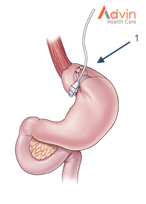
The Adjustable Gastric Band is a device made of silicone that is placed around the top part of the stomach to limit the amount of food a person can eat. It has been available in the United States since 2001. The impact on obesity related diseases and long-term weight loss is less than with other procedures. Its use has therefore declined over the past decade.

The Procedure
  1. This device is placed and secured around the top part of the stomach creating a small pouch above the band.
How it Works

The feeling of fullness depends upon the size of the opening between the pouch and the rest of the stomach. The opening size can be adjusted with fluid injections through a port underneath the skin. Food goes through the stomach normally but is limited by the smaller opening of the band. It is less successful against type 2 diabetes and has modest effects on the metabolism.

Advantages
  1. Lowest rate of complications early after surgery
  2. No division of the stomach or intestines
  3. Patients can go home on the day of surgery
  4. The band can be removed if needed
  5. Has the lowest risk for vitamin and mineral deficiencies

Single Anastomosis Duodeno-Ileal Bypass with Sleeve Gastrectomy (SADI-S)


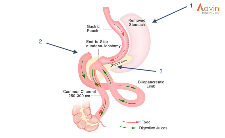
The Single Anastomosis Duodenal-Ileal Bypass with Sleeve Gastrectomy, referred to as the SADI-S is the most recent procedure to be endorsed by the American Society for Metabolic and Bariatric Surgery. While similar to the BPD-DS, the SADI-S is simpler and takes less time to perform as there is only one surgical bowel connection.

The Procedure
  1. The operation starts the same way as the sleeve gastrectomy, making a smaller tube-shaped stomach.
  2. The first part of the small intestine is divided just after the stomach.
  3. A loop of intestine is measured several feet from its end and is then connected to the stomach. This is the only intestinal connection performed in this procedure.
How it Works

When the patient eats, food goes through the pouch and directly into the latter portion of the small intestine. The food then mixes with digestive juices from the first part of the small intestine. This allows enough absorption of vitamins and minerals to maintain healthy levels of nutrition. This surgery offers good weight loss along with less hunger, more fullness, blood sugar control and diabetes improvement.

Advantages
  1. Highly effective for long-term weight loss and remission of type 2 diabetes
  2. Simpler and faster to perform (one intestinal connection) than gastric bypass or BPD-DS
  3. Excellent option for a patient who already had a sleeve gastrectomy and is seeking further weight loss

Plastic Surgery

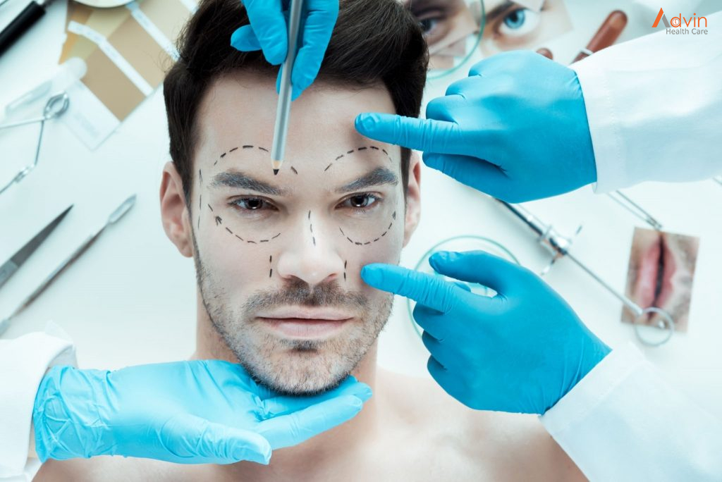
Plastic Surgery includes the repair, reconstruction, or replacement of physical defects of form or function involving the skin, musculoskeletal system, cranio-maxillofacial structures, hand, extremities, breast and trunk, and external genitalia or cosmetic enhancement to these areas of the body. Cosmetic surgery is an essential component of plastic surgery. The plastic surgeon uses cosmetic surgical procedures both to improve overall appearance and to optimize the outcome of reconstructive procedures. Competency in plastic surgery implies an amalgam of basic medical and surgical knowledge, operative judgement, technical expertise, ethical behaviour and interpersonal skills to achieve problem resolution and patient satisfaction.

History Of Plastic Surgery


Sushruta is considered the “Father of Plastic Surgery.” He lived in India sometime between 1000 and 800 BC, and is responsible for the advancement of medicine in ancient India. His teaching of anatomy, pathophysiology, and therapeutic strategies were of unparalleled luminosity, especially considering his time in the historical record. He is notably famous for nasal reconstruction, which can be traced throughout the literature from his depiction within the Vedic period of Hindu medicine to the era of Tagliacozzi during Renaissance Italy to modern-day surgical practices. The primary focus of this historical review is centered on Sushruta’s anatomical and surgical knowledge and his creation of the cheek flap for nasal reconstruction and its transition to the “Indian method.” The influential nature of the Sushruta Samhita, the compendium documenting Sushruta’s theories about medicine, is supported not only by anatomical knowledge and surgical procedural descriptions contained within its pages, but by the creative approaches that still hold true today.

What Is Plastic Surgery?


Just because the name includes the word “plastic” doesn’t mean patients who have this surgery end up with a face full of fake stuff. The name isn’t taken from the synthetic substance but from the Greek word plastikos, which means to form or mold (and which gives the material plastic its name as well).

Plastic surgery is a special type of surgery that can change a person’s appearance and ability to function.

  • Reconstructive procedures correct defects on the face or body. These include physical birth defects like cleft lips and palates and ear deformities, traumatic injuries like those from dog bites or burns, or the aftermath of disease treatments like rebuilding a woman’s breast after surgery for breast cancer.
  • Cosmetic (also called aesthetic) procedures alter a part of the body that the person is not satisfied with. Common cosmetic procedures include making the breasts larger (augmentation mammoplasty) or smaller (reduction mammoplasty), reshaping the nose (rhinoplasty), and removing pockets of fat from specific spots on the body (liposuction). Some cosmetic procedures aren’t even surgical in the way that most people think of surgery — that is, cutting and stitching. For example, the use of special lasers to remove unwanted hair and sanding skin to improve severe scarring are two such treatments.

When Plastic Surgery Is Used?


Plastic surgery can be used to repair:

  • abnormalities that have existed from birth, such as a cleft lip and palate, webbed fingers, and birthmarks
  • areas damaged by the removal of cancerous tissue, such as from the face or breast
  • extensive burns or other serious injuries

Plastic surgery can often help improve a person’s self-esteem, confidence and overall quality of life.

Plastic Surgery Techniques


Some of the techniques used during plastic surgery are:

  • Skin grafts – where healthy skin from an unaffected area of the body is removed and used to replace lost or damaged skin
  • Skin flap surgery – where a piece of tissue from one part of the body is transferred to another, along with the blood vessels that keep it alive; it’s called flap surgery because the healthy tissue usually remains partially attached to the body while it’s repositioned
  • Tissue expansion – where surrounding tissue is stretched to enable the body to “grow” extra skin, which can then be used to help reconstruct the nearby area

As well as these techniques, plastic surgeons also use many other methods, such as:

  • Fat transfer or grafting – where fat is removed from one area and inserted in another area, usually to correct unevenness
  • Vacuum closure – where suction is applied to a wound through a sterile piece of foam or gauze to draw out fluid and encourage healing
  • Camouflage make-up or cream
  • Prosthetic devices, such as artificial limbs

Advin Plastic Surgery Instrument


How Robotics Is Shaping The Future Of Urology?

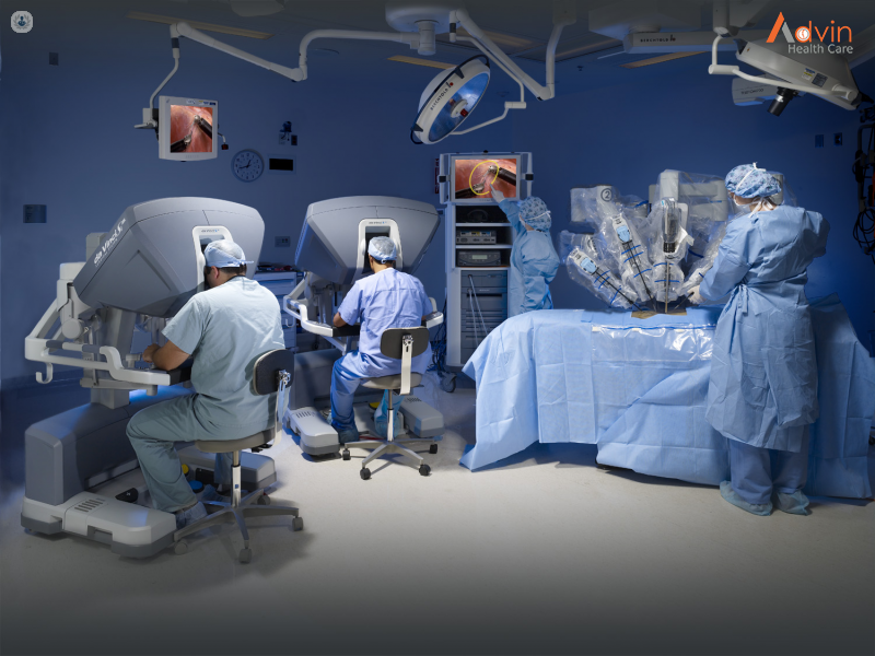
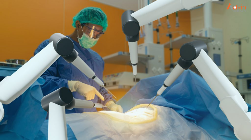
Robotic surgery or Robot-assisted surgery is a specialized technique used by Robotic Urologist to enhance the overall experience and outcome of critical surgical procedures. It makes it possible for the operating urosurgeon to reach even distant and critical spaces with a very small incision. The specialist technology also offers accurate motions and increased magnification.

The main parts of a robotic technology include:
  • Surgical arms – It comes with small tools and wrists that are used while performing the operation. This offers 7 degrees of freedom of movement
  • Specialised lens – Provides a magnified 3D image of the surgical area.
  • Surgical console – It’s kind of a dashboard for the operating surgeons to direct the movement of the device and camera.

Robotic Surgery and Urology


The enormous advancements in robotic surgery during the last ten years are expected to be surpassed by even larger advancements in the coming decade. The growing use of robotic-assisted surgical devices has propelled urology to the forefront of surgical robotics. Because accuracy is so important in pelvic procedures, urology department in Medanta has become leaders in robot-assisted surgery.

As the pelvic area is a potential space with densely packed arteries, veins, and organs, it requires a very precise intervention. Robotic surgery in urology provides several advantages that make it better than open or even laparoscopic surgery. These advantages include a decrease in the amount of blood loss, reduced patient discomfort, and minimizing the use of pain killers. It also helps in reducing the time of the hospital stay and the healing time.

What happens during Robotic Surgery?


Your operating urosurgeon will first make one or more tiny cuts as required by the procedure. Your surgeon will insert a thin tube-like device through these incisions. Instruments are put through these tubes after the robot is hooked to them. One of the holes is used to insert a long, flexible camera called an endoscope. Through the other openings, a surgical instrument that is controlled by the robotic arm is inserted.   Uro surgeon sits at a dashboard a few feet away from you, controlling the robotic arm. An assistant remains at your side to assist the surgeon by shifting the instruments as needed.

Advantages of Robotic Surgery


Robotic surgery provides several advantages over conventional surgical procedures. It provides advantages to the surgical operator as well as to the patients.

Advantages to the surgeon :
  • Greater precision: The robotic arm’s movements are more exact and provide better precision.
  • Better visualization: A high definition camera provides magnified 3D imaging that is superior to the naked eye.
  • Ability to do surgery in limited spaces inside the body
Advantages to the patients :
  • Reduced pain during recovery
  • Lower risk of infection.
  • Reduced blood loss.
  • Shorter hospital stays.
  • Smaller scars.
  • Quicker recovery time
  • Overall better clinical outcomes

Robotic Procedures in Urology


Several urology surgeries involve the use of robots. The use of the robotic procedure in urological surgeries is because of the prolonged timing of the surgical procedure, the need for steady images, and the critical location of the surgery. Laparoscopic radical prostatectomy is one such surgical procedure that is best suited for robotic surgery. Some of the common urological surgical procedures where robotic technology is utilized are:

  1. Robotic prostatectomy: It is a robot-assisted surgical procedure that removes the prostate gland
  2. Robotic cystectomy: In this procedure, the urinary bladder is removed using robotic surgery
  3. Robotic Partial Nephrectomy: The tumor bearing portion of the kidney is removed while the remaining normal kidney is repaired through small holes with robotic instruments
  4. Robotic pyeloplasty: It is a procedure used to treat a disease known as ureteropelvic junction (UPJ) obstruction.

Other procedures include


  • Nephroureterectomy
  • Adrenalectomy
  • Ureteric reimplantation
  • Kidney transplantation

Fallopian Tube Recanalization

Fallopian tube recanalization (FTR) is a therapeutic procedure performed to open an obstructed fallopian tube by passage of a guidewire and catheter through a proximal fallopian tube obstruction. The equipment and techniques used for FTR are extensions of catheter interventions used in angiography.

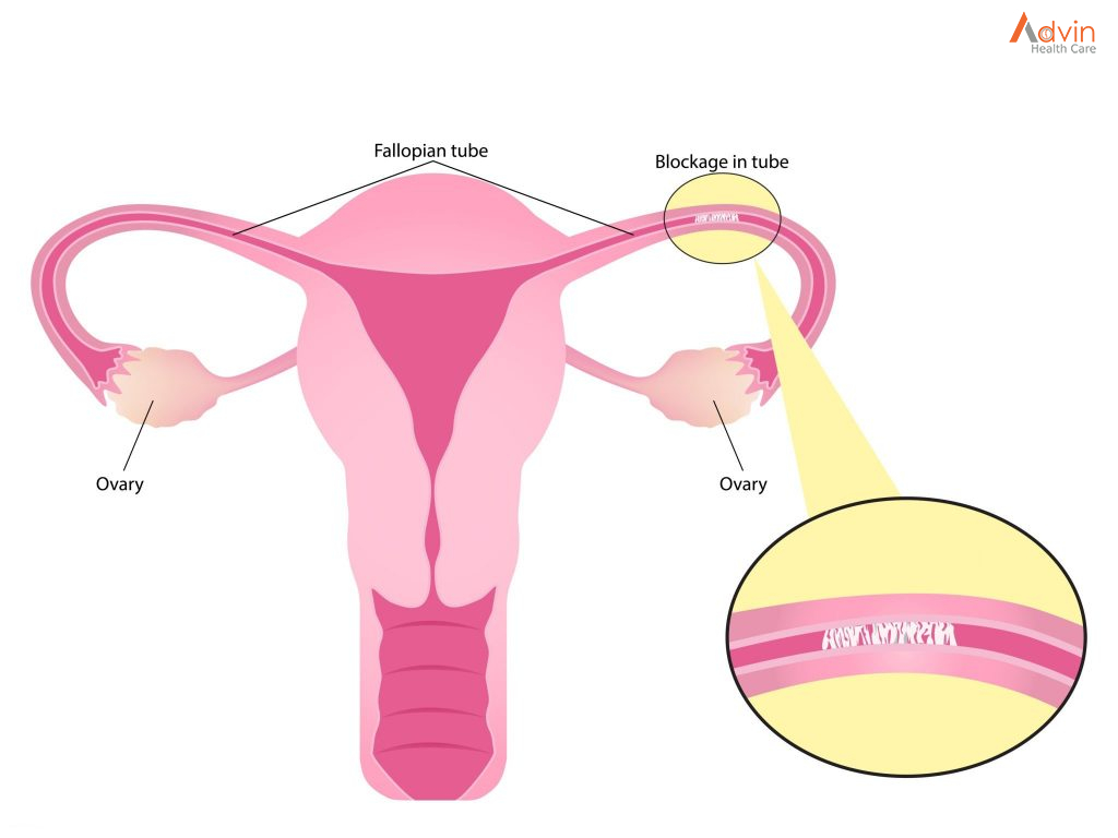
Fallopian tubes are the thin tubes in the uterus which helps in leading the mature eggs from the ovaries to the uterus. Obstruction in the Fallopian tube will prevent the egg from travelling down the tube. This condition is what is called fallopian tubal blockage which causes infertility in women. About 40% cases of infertility are caused due to this tubal blockage. It is also known as tubal factor infertility. Tubal recanalization or Fallopian tube recanalization is to remove the blockage in the fallopian tubes.

These fine tube-like structures or fallopian tubes connect the uterus and the ovaries. The eggs develop in the ovaries and travel through the fallopian tubes. They meet the sperm and fertilize in the fallopian tubes. Blockage in the tube prevents this action and whereby prevents pregnancy. Inflammation and infections of the tube is seen as the main cause for the tubal blockage. Other causes of fallopian tubal blockage can be sexually transmitted infection such as chlamydia or gonorrhea, infections caused by abortion or miscarriage, history of ectopic pregnancy, endometriosis, previous abdominal surgery etc.

Fallopian tube recanalization is all about reopening the tube to remove the blockage. During the procedure, a speculum will be placed into the vagina. A small plastic tube called a catheter will be passed through the cervix into the uterus. This procedure can be done under laparoscopic guidance or alternatively through the catheter, a liquid contrast agent will be injected. The blockage will be identified through the image shown on the screen using and X-ray camera. The fallopian tube is then opened using a small catheter threaded through the previous catheter. Through this procedure, blockages can be removed and normal function can be rebuilt.

What are fallopian tubes?


The fallopian tubes are important for female fertility. They are the passageways for the eggs to travel from the ovaries to the uterus. During conception:

  1. The ovary releases an egg, which travels into the fallopian tube.
  2. Sperm travels into the fallopian tubes to fertilize the egg.
  3. The resulting embryo is nourished and transported to the uterus where the pregnancy continues.

A common cause of female infertility is a blockage of the fallopian tubes, usually as the result of debris that has built up. Occasionally, scarring from surgery or serious infection can lead to a blockage as well.

What happens during a fallopian tube recanalization?


Fallopian tube recanalization (FTR) is a nonsurgical procedure our interventional radiologists use to treat these blockages. Recanalization is the medical term for “reopening.”

During the procedure, which does not require any needles or incisions, we will:

  1. Place a speculum into the vagina and pass a small plastic tube (catheter) through the cervix into the uterus.
  2. Inject a liquid contrast agent (sometimes called a dye, although nothing is stained) through the catheter.
  3. Examine the uterine cavity on a nearby monitor using an X-ray camera.
  4. Obtain a hystero-salpingogram or HSG. Literally, that means a “uterus-and-fallopian-tube-picture.”
  5. Determine if there is a blockage and if it is located on one or both fallopian tubes.
  6. Thread a smaller catheter through the first catheter and then into the fallopian tube to clear the blockage.

Preparation for the FTR procedure


Two days before your procedure, your gynaecologist will prescribe an antibiotic called Doxycycline to be taken twice a day, which you will continue up to and after your procedure for two more days. Since FTR is sometimes uncomfortable (though usually much less than a hysterosalpingogram, owing to much more delicate equipment being used), an intravenous line is placed prior to the procedure. Short-acting medications will be given for relaxation and pain relief. For this reason, you will be instructed to not eat anything after midnight the night before. You will also be asked to take Ibuprofen 400 mg (2 pills) the night before and the morning of your procedure.

Procedures details


Infertility is defined as the inability of a couple to conceive after one year of un- protected sex. One other common causes of infertility is blocks in the fallopian tube. Till recently this problem required microsurgery and results after surgery was never good. Over the recent years interventional radiology techniques have been extended to remove these blocks. The procedure is done under mild sedation and the patient is discharged after a few hours.

Fallopian tubes are fine little tubular structures that connect the uterus to the ovary. Once an ovum is mature it travels along the fallopian tube and it is here that fertilization takes place. Thus a block in these tubes makes fertilization impossible.

These blocks can be secondary to an infection like tuberculosis or a mucous plug. If it is a mucous plug, then removing this block can easily be achieved by passing a flexible soft wire through the blocked segment.

The procedure is normally performed on the eighth ninth or 10th day of a woman’s menstrual cycle. This is to enable pregnancy to take place in the first cycle following recanalization.

The procedure involves passing a small (sheath) tube into the uterine cavity through the vagina. This may cause mild discomfort. Through the sheath another smaller tube is passed into the uterine cavity and the opening to the fallopian tube intubated with the help of a guidewire. A brisk injection of contrast is made into the opening to flush out debris if present. If this procedure fails than the occluded segment is gently manipulated with a guidewire and the block removed.

Recanalization of these tubes is successful in close to 90% of the patients who present with cornual blocks [proximal 2 cm of the fallopian tube]. Blocks in other parts of the fallopian tube are secondary to infection and cannot be opened. It is also important to know that fallopian tubes may have disease outside usually due to adhesions. These have to be ruled out by a laparoscopy before a patient is taken up for fallopian tube recanalization.

Close to 40% of the patients who undergo successful tubal recanalization conceive within the first 6 months. However, it is important that the male partner is fully investigated and all abnormalities corrected before this procedure is taken up.

Tubes that have been recanalized may occlude again and then the procedure can be repeated.

Following the procedure, the patient may have mild cramps and little bleeding for a few days. However, all patients are encouraged to resume normal activities and have a normal sexual relationship with the husband.

Advin Products