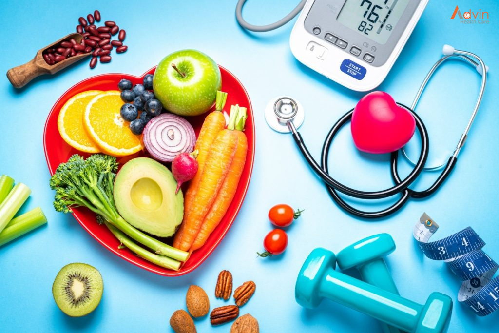
What is Nutrition?
nutritionists use ideas from molecular biology, biochemistry, and genetics to understand how nutrients affect the human body.
Nutrition also focuses on how people can use dietary choices to reduce the risk of disease, what happens if a person has too much or too little of a nutrient, and how allergies work.
Nutrients provide nourishment. Proteins, carbohydrates, fat, vitamins, minerals, fiber, and water are all nutrients. If people do not have the right balance of nutrients in their diet, their risk of developing certain health conditions increases.
Macro-nutrients
Micro-nutrients are essential in small amounts. They include vitamins and minerals. Manufacturers sometimes add these to foods. Examples include fortified cereals and rice.
Carbohydrates sugar, starch, and fiber are types of carbohydrates.
Sugars are simple carbs. The body quickly breaks down and absorbs sugars and processed starch. They can provide rapid energy, but they do not leave a person feeling full. They can also cause a spike in blood sugar levels. Frequent sugar spikes increase the risk of type 2 diabetes and its complications.
Fiber is also a carbohydrate. The body breaks down some types of fiber and uses them for energ; others are metabolized by gut bacteria, while other types pass through the body.
Fiber and unprocessed starch are complex carbs. It takes the body some time to break down and absorb complex carbs. After eating fiber, a person will feel full for longer. Fiber may also reduce the risk of diabetes, cardiovascular disease, and colorectal cancer. Complex carbs are a more healthful choice than sugars and refined carbs.
Proteins
Proteins consist of amino acids, which are organic compounds that occur naturally.
There are 20 amino acids. Some of these are essential Trusted Source, which means people need to obtain them from food. The body can make the others.
Some foods provide complete protein, which means they contain all the essential amino acids the body needs. Other foods contain various combinations of amino acids.
Most plant-based foods do not contain complete protein, so a person who follows a vegan diet needs to eat a range of foods throughout the day that provides the essential amino acids.
Fats
Fats are essential for:
- lubricating joints
- helping organs produce hormones
- enabling the body to absorb certain vitamins
- reducing inflammation
- preserving brain health
Too much fat can lead to obesity, high cholesterol, liver disease, and other health problems.
However, the type of fat a person eats makes a difference. Unsaturated fats, such as olive oil, are more healthful than saturated fats, which tend to come from animals.
Water
The adult human body is up to 60% water, and it needs water for many processes. Water contains no calories, and it does not provide energy.
Many people recommend consuming 2 liters, or 8 glasses, of water a day, but it can also come from dietary sources, such as fruit and vegetables. Adequate hydration will result in pale yellow urine.
Requirements will also depend on an individual’s body size and age, environmental factors, activity levels, health status, and so on.
Minerals
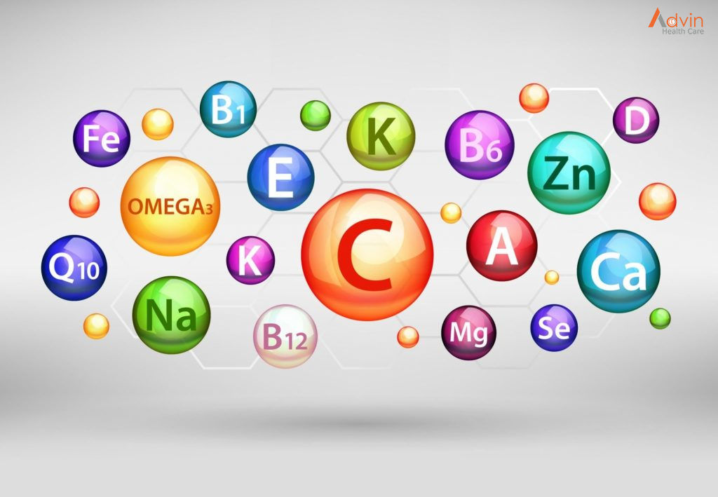
The body needs carbon, hydrogen, oxygen, and nitrogen. It also needs dietary minerals, such as iron, potassium, and so on. In most cases, a varied and balanced diet will provide the minerals a person needs. If a deficiency occurs, a doctor may recommend supplements. Here are some of the minerals the body needs to function well.
Potassium
Potassium is an electrolyte. It enables the kidneys, the heart, the muscles, and the nerves to work properly. Too little can lead to high blood pressure, stroke, and kidney stones.Too much may be harmful to people with kidney disease. Avocados, coconut water, bananas, dried fruit, squash, beans, and lentils are good sources.
Sodium
Sodium is an electrolyte that helps:
- maintain nerve and muscle function
- regulate fluid levels in the body
Too little can lead to hyponatremia. Symptoms include lethargy, confusion, and fatigue. Too much can lead to high blood pressure, which increases the risk of cardiovascular disease and stroke. Table salt, which is made up of sodium and chloride, is a popular condiment. However, most people consume too much sodium, as it already occurs naturally in most foods. Experts urge people not to add table salt to their diet. Current guidelines recommend consuming no more than 2,300 mg of sodium a day, or around one teaspoon. This recommendation includes both naturally-occurring sources, as well as salt a person adds to their food. People with high blood pressure or kidney disease should eat less.
Calcium
The body needs calcium Trusted Source to form bones and teeth. It also supports the nervous system, cardiovascular health, and other functions.
Too little can cause bones and teeth to weaken. Symptoms of a severe deficiency include tingling in the fingers and changes in heart rhythm, which can be life-threatening. Too much can lead to constipation, kidney stones, and reduced absorption of other minerals. Current guidelines for adults recommend consuming 1,000 mg a day, and 1,200 mg for women aged 51 and over. Good sources include dairy products, tofu, legumes, and green, leafy vegetables.
Phosphorus
Phosphorus is present in all body cells and contributes to Trusted Source the health of the bones and teeth. Too little phosphorus can lead to bone diseases, affect appetite, muscle strength, and coordination. It can also result in anemia, a higher risk of infection, burning or prickling sensations in the skin, and confusion. Too much in the diet is unlikely to cause health problems though toxicity is possible from supplements, medications, and phosphorus metabolism problems. Adults should aim to consume around 700 mgTrusted Source of phosphorus each day. Good sources include dairy products, salmon, lentils, and cashews.
Magnesium
Magnesium contributes to Trusted Source muscle and nerve function. It helps regulate blood pressure and blood sugar levels, and it enables the body to produce proteins, bone, and DNA. Too little magnesium can eventually lead to weakness, nausea, tiredness, restless legs, sleep conditions, and other symptoms. Too much can result in digestive and, eventually, heart problems. Nuts, spinach, and beans are good sources of magnesium. Adult females need 320 mg Trusted Source of magnesium each day, and adult males need 420 mg.
Zinc
Zinc plays a role in the health of body cells, the immune system, wound healing, and the creation of proteins. Too little can lead to hair loss, skin sores, changes in taste or smell,and diarrhea, but this is rare. Too much can lead to digestive problems and headaches. Adult females need 8 mg Trusted Source of zinc a day, and adult males need 11 mg. Dietary sources include oysters, beef, fortified breakfast cereals, and baked beans. For more on dietary sources of zinc,
Iron
Iron is crucial for the formation Trusted Source of red blood cells, which carry oxygen to all parts of the body. It also plays a role in forming connective tissue and creating hormones. Too little can result in anemia, including digestive issues, weakness, and difficulty thinking. Learn more here about iron deficiency. Too much can lead to digestive problems, and very high levels can be fatal. Good sources include fortified cereals, beef liver, lentils, spinach, and tofu. Adults need 8 mg Trusted Source of iron a day, but females need 18 mg during their reproductive years.
Manganese
The body uses manganese to produce energy Trusted Source, it plays a role in blood clotting, and it supports the immune system. Too little can result in weak bones in children, skin rashes in men, and mood changes in women. Too much can lead to tremors, muscle spasms, and other symptoms, but only with very high amounts. Mussels, hazelnuts, brown rice, chickpeas, and spinach all provide manganese. Male adults need 2.3 mg Trusted Source of manganese each day, and females need 1.8 mg.
Copper
Copper helps the body Trusted Source make energy and produce connective tissues and blood vessels. Too little copper can lead to tiredness, patches of light skin, high cholesterol, and connective tissue disorders. This is rare. Too much copper can result in liver damage, abdominal pain, nausea, and diarrhea. Too much copper also reduces the absorption of zinc. Good sources include beef liver, oysters, potatoes, mushrooms, sesame seeds, and sunflower seeds. Adults need 900 microgramsTrusted Source (mcg) of copper each day.
Selenium
Selenium is made up of over 24 selenoproteins, and it plays a crucial roleTrusted Source in reproductive and thyroid health. As an antioxidant, it can also prevent cell damage. Too much selenium can cause garlic breath, diarrhea, irritability, skin rashes, brittle hair or nails, and other symptoms. Too little can result in heart disease, infertility in men, and arthritis. Adults need 55 mcg Trusted Source of selenium a day.