Diagnostic Radiology
Radiology, also known as diagnostic imaging, is a series of tests that take pictures or images of parts of the body. The field encompasses two areas — diagnostic radiology and interventional radiology — that both use radiant energy to diagnose and treat diseases. While there are several different imaging exams, some of the most common include x-ray, MRI, ultrasound, CT scan and PET scan.
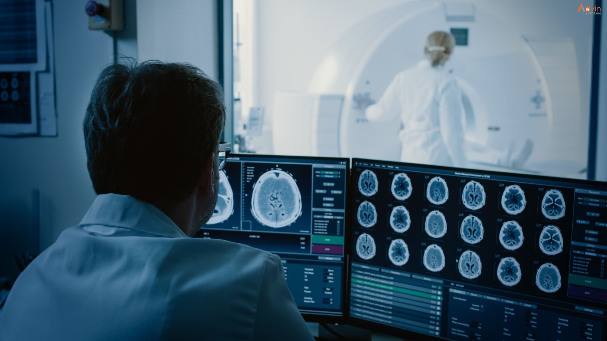
A radiologist will look at the outcome of a certain imaging test to find a relevant image that evaluates and supports a diagnosis. These individuals are usually medical doctors (MDs) with highly specialized training focused on the interpretation of medical imaging. Radiologic technologists also aid in this process, as they use and manage the machines in the course of producing an image. After a patient undergoes imaging tests, radiologists will give reports of their interpretations to the referring clinical doctors.
History of Radiology
Radiology began in Germany in 1895 when Wilhelm Conrad Röntgen made an energized, lightproof cathode ray tube that started to fluoresce when situated a couple of feet away from a screen painted with fluorescent material. He knew the screen was responding to unknown rays transmitted throughout the room, which he called “x-rays.” After Röntgen’s discovery, people began creating radiographic images that started as a burst of ionizing radiation and created a contrast image on a piece of film.
What Is Radiology Used for?
Radiology is used for a wide range of conditions, and is classified depending on the type of radiology and the exact imaging test used. The various imaging exams include:
- Radiographs: X-rays to look at bones, the chest or the abdomen.
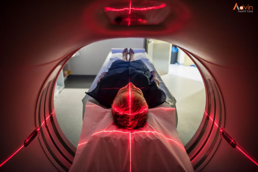
- CT (Computed Tomography): A CT captures multiple x-ray angles of the patient using a doughnut-shaped machine, then creates computer-processed images.
- MRI (Magnetic Resonance Imaging): An MRI uses magnetic fields and radio waves with computer processing to create images.
- Mammograms: Specially powered x-rays that look at breast tissues.
- Ultrasound: An ultrasound uses sound waves to create moving images that display on a monitor, commonly used for echocardiograms and examining the womb during pregnancy.
- Fluoroscopy: X-rays that make moving images of the body in real time. This imaging is crucial for many procedures, especially those involving the gastrointestinal tract.
- Nuclear medicine: These are short-acting radioactive substances that generate light from bodily processes. A camera collects the light, so a computer can process it and develop an image.
Diagnostic radiology uses these imaging results to identify a wide range of problems, from broken bones to heart conditions and blood clots. Interventional radiology also uses imaging such as CT scans, MRI and ultrasounds to guide medical procedures. Patients are typically awake during these procedures, whether it’s treating cancer, back pain, or liver and kidney problems. In some cases, interventional radiology eliminates the need for surgery and scopes.
Diagnostic Imaging
Diagnostic radiology refers to the field of medicine that uses non-invasive imaging scans to diagnose a patient. The tests and equipment used sometimes involves low doses of radiation to create highly detailed images of an area.
Examples of diagnostic radiology include:
- Radiography (X-rays)
- Ultrasound
- Computed Tomography (CT) Scans
- Magnetic Resonance Imaging (MRI) Scans
- Nuclear Medicine Scans
Diagnostic radiology can be used to identify a wide range of problems. Broken bones, heart conditions, blood clots, and gastrointestinal conditions are just a few of the problems that can be identified by diagnostic imaging.
In addition to identifying problems, doctors can use diagnostic radiology to monitor how your body is responding to a current treatment. Diagnostic radiology can also screen for diseases such as breast cancer and colon cancer.
Technology Used in Radiology
The technology and machinery used in radiology varies from method to method. Some use radiation while others do not.
The most common machines used in radiology are:
- X-ray Machine: Uses X-rays, a type of electromagnetic radiation, to produce images of the interior of the body without having to make any incisions.
- CT Scanner: Uses X-ray equipment to create a sequence of cross-sectional images of the body. Often used when a doctor needs highly detailed images to study in order to identify the source of a problem, especially on soft tissue.
- MRI Machine: Uses a magnetic field instead of radiation to produce images of the inside of a body. Used for parts of the body that CT scanners cannot produce clear images of, such as bones.
Some of the diagnostic tests may require compounds to be ingested or chemicals to be injected for a clear view of your blood veins. Other tests may require anaesthesia and a scope in order for a doctor to clearly determine the problem.
Interventional Radiology
Interventional radiology uses imaging technology such as CT scans, MRI, and Ultrasounds to help guide medical procedures. This technology eliminates the need for surgery and scopes to diagnose and treat certain conditions. Instead, patients are often awake during the procedure or under very mild sedation.
Common uses for interventional radiology include:
- Treating cancers
- Treating blockages in arteries or veins
- Treating back pain
- Treating liver and kidney problems
Interventional radiologists are highly specialized radiologists who have extra training and experience to safely and accurately perform these procedures.
Advin Radiology Product

