Radiology defines a wing of medicine that functions using radiant energy while diagnosing and treating diseases. In simple words, radiology means a process to determine whether a medical condition is present or not before moving ahead with the treatment. It is a test that takes images of different body parts and tries to find relevant issues to support a necessary diagnosis.
This branch of medicine comprises two areas – diagnostic radiology and interventional radiology. Both these forms use different radiant energy to provide a required diagnosis. Among the various radio imaging exams, some of the most common include- MRI, Ultrasound, X-ray, PET scan, etc. After the patients experience these tests, the radiologist will provide reports of their elucidations to the concerned doctors.
Now that individuals have a fair understanding of what radiology is, they should know about this diagnostic test’s different types and procedures.
The five most common radiological tests prescribed by doctors are explained below.
X-Rays
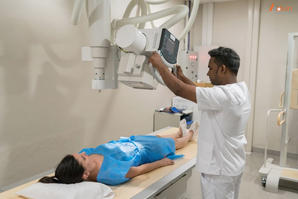
X-ray is the most common and widely performed radiological test. It is non-invasive and painless. This method makes use of electromagnetic radiation to detect even minor anomalies. Modern x-ray machines have become more efficient, with a ray source on one side and radiation-sensitive plates on the other. It helps in quick imaging and gives instant results.
The Test Procedure
The patient has to place the concerned body part between the ray source and the radiation plate. The doctor might ask them to move or turn to take images from different angles. This process takes only about 10 to 15 minutes.
Simple x-rays are done for screening as well as diagnostic purposes. It can:
- detect hairline fractures, ligament tears, or lumps in certain areas
- diagnose conditions like osteoporosis, arthritis, digestive tract issues, and infections
- track the severity of an injury and the effects of treatment on the patient’s body
- screening for and diagnosing the malignancy of breast lumps (mammogram)
- live imaging of blood flow, hard and soft tissues, and joints using a contrast dye like barium (fluoroscopy/barium swallow radiology)
Ultrasound
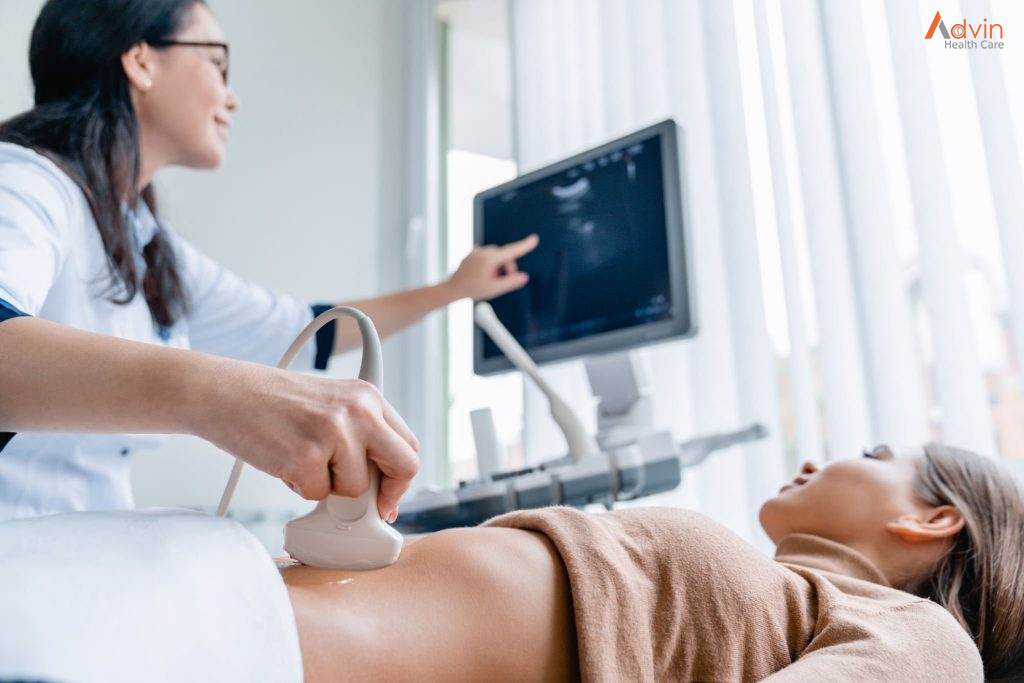
Ultrasound, also called sonography, is another widespread radiological procedure. It uses high-frequency sound waves to detect different structures inside the body. There are no radiation waves in this test which makes this a safe procedure even for pregnant women. Ultrasound can be minimally invasive or non-invasive and mainly studies soft tissue structures and organs.
The Test Procedure
The doctor applies a gel on the target body part and places a transducer or probe which emits ultrasound waves. They move the probe around the body part to get a clear picture. For endoscopic ultrasound, the probe is inserted into the body through the patient’s mouth or rectum, depending on the part to be studied. The procedure can take about 25 minutes to 1 hour.
Purpose Of The Test
Ultrasound is used for diagnostic and interventional purposes. It is performed to:
- diagnose breast lumps, joint inflammation, and issues related to gall bladder and prostate
- diagnose inflammation or cancer in the digestive tract
- study the progression of a pregnancy
- act as a guide during biopsies
CT/CAT Scan
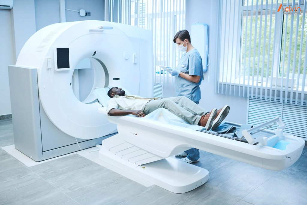
A computerised Axial Tomography scan uses a series of ionising radiations to form a cross-sectional image of the patient’s whole body. It is a non-invasive procedure and helps study almost all body structures.
The Test Procedure
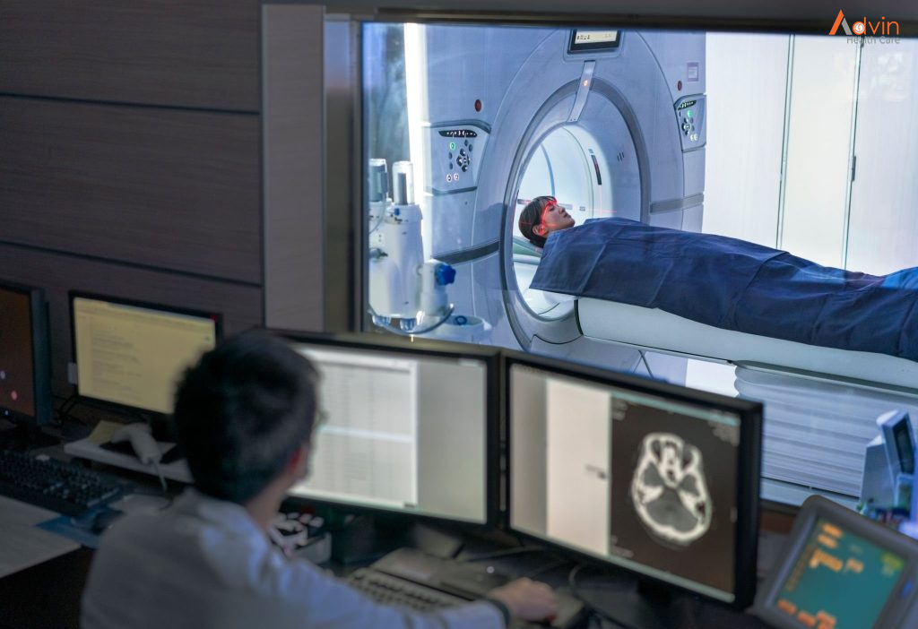
The patient is made to lie on a table that slides into the doughnut-shaped scanner. A scanning tube at the centre rotates around the patient’s body and creates a 3-D image. There is a transmitter inside the scanner through which the patient can talk to the doctor in case of nervousness or discomfort. The imaging takes only 15 to 20 minutes.
Purpose Of The Test
Doctors usually prescribe a CAT scan when other tests indicate some abnormality. It is done for diagnostic and radiology treatment purposes like:
- studying the severity of a traumatic injury or fracture
- detecting tumours and other abnormalities
- diagnosing infections and heart and vascular diseases
- guiding biopsies
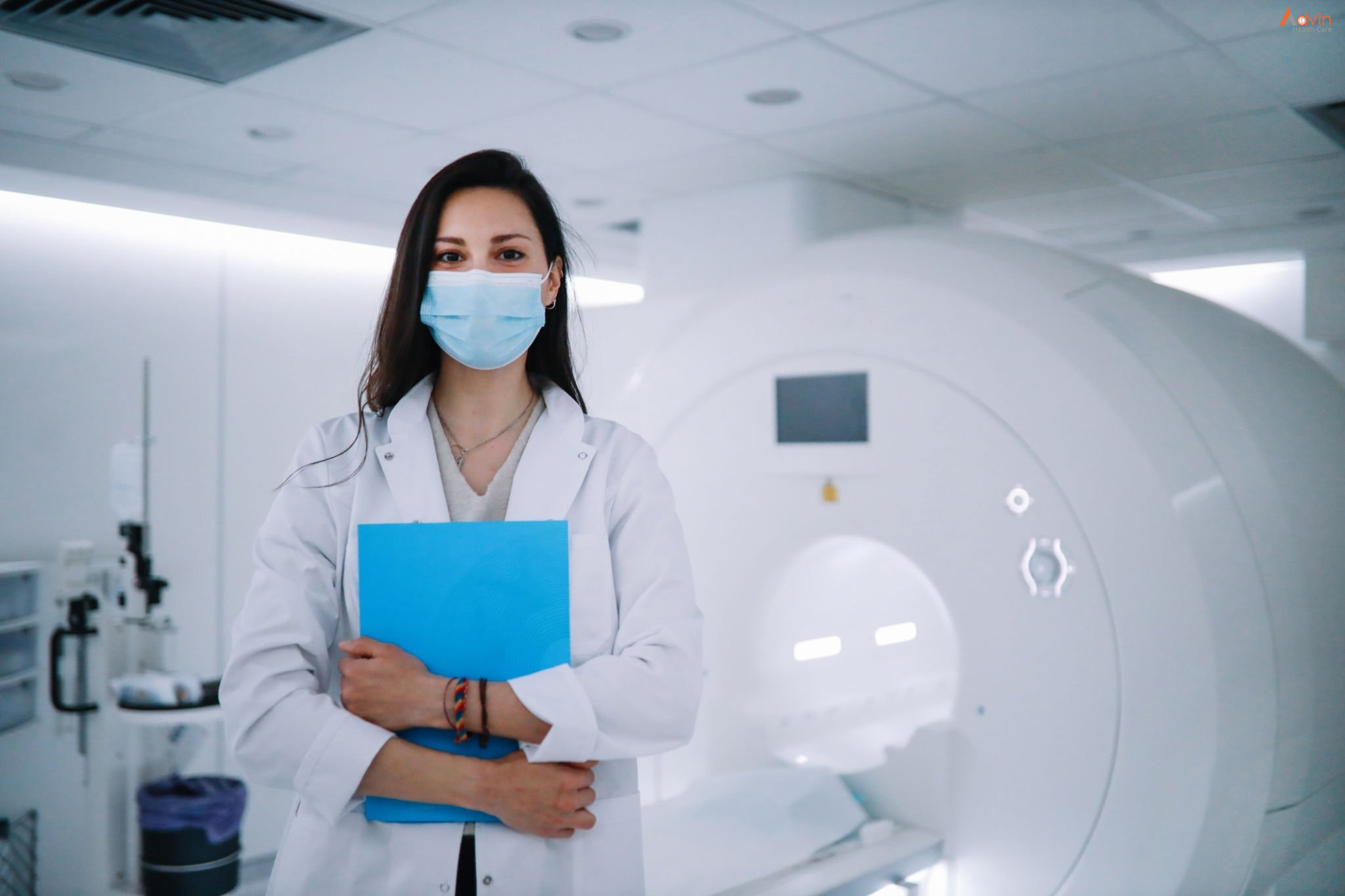
MRI Scan
Magnetic Resonance Imaging works very similarly to a CAT scan. Only, it uses magnetic fields instead of ionising radiation. This test works for almost all body parts and conditions. Due to the absence of radiation, it is also considered a safer method. However, it takes longer than a CAT scan.
The Test Procedure
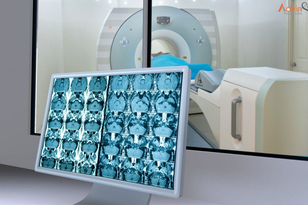
The patient is made to lie on the sliding table, which moves into the MRI scanner. The scanner rotates, sending magnetic waves to create a 3-D image of the patient’s body. As it does so, the scanner makes loud noises. The patient can interact with the doctor through the transmitter in case of discomfort.
Purpose Of The Test
MRI scan helps in diagnosing a wide array of conditions. It includes:
- spinal cord diseases
- issues related to soft tissues like organs, blood vessels, joints, and tendons
- strokes, aneurysms, and multiple sclerosis
PET Scan
Positron Emission Tomography scan follows a different methodology than the ones discussed above. In this test, a radioactive drug, called a radiotracer, is inserted into the patient’s body. The PET scanner studies the transmission of this tracer on a cellular level. This radiography test helps study any abnormalities and the functioning of different body systems.
The Test Procedure
The radiotracers are introduced into the patient’s body via an injection into the veins or inhaling gases, or drinking a mixture containing radiotracers. It depends on the organ system or body part to be studied. After about an hour of introducing the radiotracer into the body, the scanning is done. The whole procedure might take up to 2 hours. The radiotracers are introduced into the patient’s body via an injection into the veins, inhaling gases, or drinking a mixture containing radiotracers.
Purpose Of The Test
PET scan is a highly efficient procedure for performing diagnostic imaging. It is used to diagnose:
- cancer
- cardiovascular diseases
- Alzheimer’s disease
- Parkinson’s disease
- digestive tract diseases
- seizures
- epilepsy

