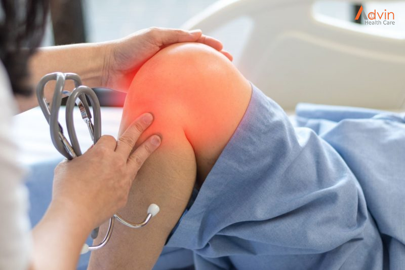
Knee Arthroscopy

Knee Arthroscopy


Knee arthroscopy is a surgical procedure that allows doctors to view the knee joint without making a large incision (cut) through the skin and other soft tissues. Arthroscopy is used to diagnose and treat a wide range of knee problems.

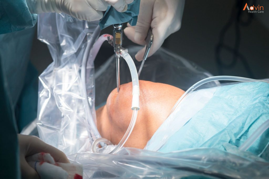
During knee arthroscopy, your surgeon inserts a small camera, called an arthroscope, into your knee joint. The camera displays pictures on a video monitor, and your surgeon uses these images to guide miniature surgical instruments.

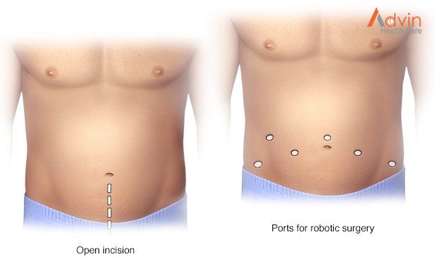
Because the arthroscope and surgical instruments are thin, your surgeon can use very small incisions, rather than the larger incision needed for open surgery. This results in less pain and joint stiffness for patients, and often shortens the time it takes to recover and return to favourite activities.

Your knee is the largest joint in your body and one of the most complex. The bones that make up the knee include the lower end of the femur (thighbone), the upper end of the tibia (shinbone), and the patella (kneecap).

Other important structures that make up the knee joint include:

  • Articular cartilage.The ends of the femur and tibia, and the back of the patella are covered with articular cartilage. This slippery substance helps your knee bones glide smoothly across each other as you bend or straighten your leg.
  • The knee joint is surrounded by a thin lining called synovium. This lining releases a fluid that lubricates the cartilage and reduces friction during movement.
  • Two wedge-shaped pieces of meniscal cartilage between the femur and tibia act as shock absorbers. Different from articular cartilage, the meniscus is tough and rubbery to help cushion and stabilize the joint.
  • Bones are connected to other bones by ligaments. The four main ligaments in your knee act like strong ropes to hold the bones together and keep your knee stable.
    • The two collateral ligaments are found on either side of your knee.
    • The two cruciate ligaments are found inside your knee joint. They cross each other to form an X with the anterior cruciate ligament in front and the posterior cruciate ligament in back.

Who needs knee arthroscopy?


Your healthcare provider may recommend knee arthroscopy if you have knee pain that doesn’t get better with nonsurgical treatments. Nonsurgical treatments include rest, ice, nonsteroidal anti-inflammatory drugs and physical therapy (PT). Although arthritis causes knee pain, arthroscopic knee surgery isn’t always an effective treatment for osteoarthritis.

Healthcare providers use arthroscopy to get a better look at cartilage, bones and soft tissues inside of your knee. They use the procedure to diagnose several types of knee injuries. Most of these injuries affect ligaments and cartilage in your knee joint.

Knee injuries among athletes (including adolescents) are very common. They can happen in contact sports and those that require jumping, such as volleyball.

What happens during knee arthroscopy?


During the procedure, your healthcare provider:

  1. Cleans your leg and secures your knee in a stabilizing device. The device ensures that your knee stays in the proper position throughout the procedure.
  2. Makes a small incision (cut) in your knee and inserts a long metal tool called an arthroscope into the incision. The arthroscope has a camera on the end. Images from the camera appear on a screen in the operating room.
  3. Looks at the images on the monitor and uses them to diagnose injuries and guide the procedure. If you need surgery, your healthcare provider makes other incisions in your knee and inserts tiny tools through them.
  4. Repairs torn tissues, shaves off damaged bone or cartilage and removes inflamed or damaged tissues. Your healthcare provider uses specially designed tools for these tasks.
  5. Closes the incisions with stitches or small bandages, and wraps your knee with a larger bandage or dressing.

What are the advantages of knee arthroscopy?


Minimally invasive procedures like knee arthroscopy usually require less recovery time than traditional (open) surgery. As you only need a few small stitches, you’re more likely to get back on your feet more quickly than with traditional surgery. You may also have less pain and a lower risk of infection.

Advin Arthroscopy Instrument


Glaucoma Surgery

Glaucoma


Glaucoma is a group of eye conditions that damage the optic nerve. The optic nerve sends visual information from your eye to your brain and is vital for good vision. Damage to the optic nerve is often related to high pressure in your eye. But glaucoma can happen even with normal eye pressure.

Glaucoma can occur at any age but is more common in older adults. It is one of the leading causes of blindness for people over the age of 60.

Many forms of glaucoma have no warning signs. The effect is so gradual that you may not notice a change in vision until the condition is in its later stages.

It’s important to have regular eye exams that include measurements of your eye pressure. If glaucoma is recognized early, vision loss can be slowed or prevented. If you have glaucoma, you’ll need treatment or monitoring for the rest of your life.

5 Types of Glaucoma Surgery


The following are types of glaucoma surgeries proven to restore normal eye pressure:

1. Laser surgery

Laser therapy is a common treatment for glaucoma. For instance, selective laser trabeculoplasty (SLT) is a first-line treatment for open-angle glaucoma.3

During SLT surgery, an ophthalmologist uses a laser beam to make microscopic holes in the eye that enable eye fluid to flow better.

Other types of laser surgeries for glaucoma include argon laser trabeculoplasty (ALT) and laser peripheral iridotomy (LPI)

According to the Glaucoma Research Foundation (GRF), laser glaucoma surgery may reduce intraocular pressure by 20 to 30%. It’s effective in roughly 80% of patients.4

2. Electrocautery

Electrocautery is a minimally invasive glaucoma surgery (MIGS).5

During this procedure, the surgeon will create a tiny incision in the eye’s drainage tubes using a device known as a trabectome.

The device uses heat to remove tissue in the eye, releasing the built-up fluid.

According to studies, trabectome surgery can reduce eye pressure by 30 percent.6

3. Trabeculectomy

Trabeculectomy is also effective in reducing IOP.

During trabeculectomy, the surgeon will create a small flap in the white area of the affected eye. This creates a drainage pathway for excess fluid.

You may need medication after the surgery to prevent scar tissue from forming.

4. Drainage Implant Surgery

Drainage implant surgery is an invasive surgical procedure.

It involves the placement of a small drainage tube known as an aqueous shunt or tube shunt in the affected eye to drain the fluid.7

Glaucoma drainage implant surgery is performed in a hospital or an outpatient surgical facility. It usually takes about an hour or less to perform the procedure.

5. Microtrabeculectomy

Microtrabeculectomy involves inserting microscopic-sized tubes into the drainage angle to drain excess aqueous fluid.

The fluid drains from the anterior chamber of the eye to beneath the conjunctiva (the eye’s outer membrane).

What to Expect Before & After Surgery


Glaucoma surgery is done at your doctor’s office or an outpatient not facility.

Below is what to expect before and after surgery:

Before


Before undergoing surgery, the medical staff will prepare you mentally to ensure you’re comfortable. You will then go to the operation room, where you will lie on the operating table.

In the room, there will be an eye surgeon, a nurse, and surgical assistants.

Your eyes will be cleaned and your face covered, leaving only the infected eye exposed.

Your doctor will also install devices to monitor your heart rate, blood pressure, and oxygen levels throughout the procedure.

An anesthesiologist will also be present to administer sedatives to keep you relaxed during surgery.

After

After surgery, you’ll be allowed to rest your eyes for a moment.

During this time, your eye doctor will continuously monitor your eye pressure. If everything seems okay, you’ll be discharged.

However, before you leave, the eye doctor will instruct you on the dos and don’ts after glaucoma surgery.

You may also be required to schedule a follow-up appointment.

After glaucoma surgery, your eye will be patched up for some time. This may affect your ability to see. In addition, the effects of sedation also affect your ability to operate machinery.

To be safe, make sure you have someone to drive you home.

What are the benefits of surgery?


The aim is to lower the pressure in your eye to reduce the risk of further damage to the optic nerve.

Advin Instruments Used In Glaucoma Surgery


Retinal Detachment Surgery

Retinal Detachment Surgery


Retinal detachment describes an emergency situation in which a thin layer of tissue (the retina) at the back of the eye pulls away from its normal position.

Retinal detachment separates the retinal cells from the layer of blood vessels that provides oxygen and nourishment to the eye. The longer retinal detachment goes untreated, the greater your risk of permanent vision loss in the affected eye.

Warning signs of retinal detachment may include one or all of the following: reduced vision and the sudden appearance of floaters and flashes of light. Contacting an eye specialist (ophthalmologist) right away can help save your vision.

What is a Retinal Detachment?


The retina is the light-sensitive layer of nerve tissue that lines the inside of the eye and sends visual messages through the optic nerve to the brain. A retinal detachment occurs when the retina becomes separated from the rest of the layers of the eye. This usually occurs after you develop a tear in the retina. The extent of permanent damage depends on how much of the retina becomes detached and whether or not the centre of the retina (the macula) becomes detached. The macula is made up of special nerve cells that provide the sharp central vision needed for seeing fine detail. If your macula has become detached, you have a poorer visual prognosis and you may not regain good enough vision to read or drive with that eye even after successful surgery.

Why do I have a Retinal Detachment? What are the symptoms?


A retinal detachment occurs when a tear forms in the retina allowing fluid to get under the retina forming a detachment. They are more common in patients who are very near- sighted, have a family history of retinal detachment, and in eyes that have had prior trauma or eye surgery. Patients often complain of flashes, new floaters and a shadow forming in their vision when a retinal detachment occurs.

Surgical Procedure


Your retinal detachment surgery will likely involve a scleral buckling and/or vitrectomy procedure. We use the most advanced surgical equipment and techniques available for retinal detachment surgery. A scleral buckling surgery involves positioning a silicone band around your eye beneath your eye muscles to bring in the walls of your eye. This elongates your eye and makes you more near sighted. A vitrectomy surgery involves making 3 holes in the eye and using instruments to remove the jelly-like substance (the vitreous humour) that normally fills the centre of the eye. The removal of the vitreous inside the eye does not cause any permanent harm. The vitreous is replaced by natural fluid produced inside the eye. The retina is then reattached and all retinal tears surrounded by laser. The eye is then filled with an inert gas to keep the retina in position as it heals. The gas bubble will dissipate from your eye within 4-6 weeks. You cannot change elevation (fly on an airplane) or undergo general anaesthesia with nitrous oxide gas while a gas bubble is in your eye. We will place a green bracelet around your wrist indicating this after surgery, do not take off the bracelet until the gas dissipates from your eye. In certain cases, we may use silicone oil instead of gas; your surgeon will review with you if this is appropriate for your surgery.

Retinal reattachment surgery usually takes one-two hours to perform. It is typically performed the under local anaesthesia so that you are awake and comfortable during the procedure and have minimal complications from anaesthesia postoperatively. If you are awake, it is very important for you stay still during surgery.

What are the benefits of Retina surgery?


The clear benefit of retinal detachment surgery is that it prevents you from blindness in that eye. The degree of recovery of vision is dependent upon a multitude of factors. These include the initial cause of retinal detachment, extent of retinal detachment by the time of initial surgery, pre-existing eye conditions, the presence of retinal PVR scarring, whether your retina re–detaches, and the success of initial surgery which is significantly dependent upon the experience and competence of your surgeon.

Advin Instrument For Retina Surgery


Appendectomy Surgery

Appendectomy


Appendicitis is an inflammation of the appendix, a finger-shaped pouch that projects from your colon on the lower right side of your abdomen.

Appendicitis causes pain in your lower right abdomen. However, in most people, pain begins around the navel and then moves. As inflammation worsens, appendicitis pain typically increases and eventually becomes severe.

Although anyone can develop appendicitis, most often it occurs in people between the ages of 10 and 30. Standard treatment is surgical removal of the appendix.

What Is an Appendectomy?


An appendectomy is the surgical removal of the appendix. It’s a common emergency surgery that’s performed to treat appendicitis, an inflammatory condition of the appendix.

The appendix is a small, tube-shaped pouch attached to your large intestine. It’s located in the lower right side of your abdomen. The exact purpose of the appendix isn’t known. However, it’s believed that it may help us recover from diarrhea, inflammation, and infections of the small and large intestines. These may sound like important functions, but the body can still function properly without an appendix.

When the appendix becomes inflamed and swollen, bacteria can quickly multiply inside the organ and lead to the formation of pus. This buildup of bacteria and pus can cause pain around the belly button that spreads to the lower right section of the abdomen. Walking or coughing can make the pain worse. You may also experience nausea, vomiting, and diarrhea.

It’s important to seek treatment right away if you’re having symptoms of appendicitis. When the condition goes untreated, the appendix can burst (perforated appendix) and release bacteria and other harmful substances into the abdominal cavity. This can be life-threatening, and will lead to a longer hospital stay.

Appendectomy is the standard treatment for appendicitis. It’s crucial to remove the appendix right away, before the appendix can rupture. Once an appendectomy is performed, most people recover quickly and without complications.

There are 2 types of surgery to remove the appendix. The standard method is an open appendectomy. A newer, less invasive method is a laparoscopic appendectomy.

  • Open appendectomy. A cut or incision about 2 to 4 inches long is made in the lower right-hand side of your belly or abdomen. The appendix is taken out through the incision.
  • Laparoscopic appendectomy. This method is less invasive. That means it’s done without a large incision. Instead, from 1 to 3 tiny cuts are made. A long, thin tube called a laparoscope is put into one of the incisions. It has a tiny video camera and surgical tools. The surgeon looks at a TV monitor to see inside your abdomen and guide the tools. The appendix is removed through one of the incisions.

During a laparoscopic surgery, your provider may decide that an open appendectomy is needed.

If your appendix has burst and infection has spread, you may need an open appendectomy.

A laparoscopic appendectomy may cause less pain and scarring than an open appendectomy. For either type of surgery, the scar is often hard to see once it has healed.

Both types of surgery have low risk of complications. A laparoscopic appendectomy has a shorter hospital stay, shorter recovery time, and lower infection rates.

Recently, some studies have suggested that intravenous antibiotics alone could cure appendicitis without the need for appendectomy. These results remain controversial and appendectomy remains the standard of care.

What happens during an appendectomy?


In most cases an appendectomy is an emergency surgery and will require a hospital stay. You will have either an open appendectomy or a laparoscopic appendectomy. This will depend on your condition and your healthcare provider’s practices.

An appendectomy is done while you are given medicines to put you into a deep sleep (under general anesthesia).

Generally, the appendectomy follows this process:

  • You will be asked to remove any jewelry or other objects that might get in the way during surgery.
  • You will be asked to remove your clothing and will be given a gown to wear.
  • An IV (intravenous) line will be put in your arm or hand.
  • You will be placed on the operating table on your back.
  • If there is a lot of hair at the surgical site, it may be clipped off.
  • A tube will be put down your throat to help you breathe. The anesthesiologist will check your heart rate, blood pressure, breathing, and blood oxygen level during the surgery.

Open appendectomy


  • A cut or incision will be made in the lower right part of your belly.
  • Your abdominal muscles will be separated and the abdominal area will be opened.
  • Your appendix will be tied off with stitches and removed.
  • If your appendix has burst or ruptured, your abdomen will be washed out with salt water (saline).
  • The lining of your abdomen and your abdominal muscles will be closed with stitches. A small tube may be put in the incision to drain out fluids.

Laparoscopic appendectomy


  • A tiny incision will be made for the tube (laparoscope). More cuts may be made so that other tools can be used during surgery.
  • Carbon dioxide gas will be used to swell up your abdomen so that your appendix and other organs can be easily seen.
  • The laparoscope will be put in and your appendix will be found.
  • Your appendix will be tied off with stitches and removed through an incision.
  • When the surgery is done, the laparoscope and tools will be removed. The carbon dioxide will be let out through the cuts. A small tube may be placed in the cut to drain out fluids.

Advantages of appendectomy


Results may vary depending upon the type of procedure and patient’s overall condition. Common advantages are:

  • Less postoperative pain
  • May shorten hospital stay
  • May result in a quicker return to bowel function
  • Quicker return to normal activity
  • Better cosmetic results

Advin Instrument Use In Appendectomy Surgery


Kirschner wires

Kirschner wires (K-wires)


Kirschner wires (K-wires) are stiff, straight wires that are sometimes needed to repair a fracture (broken bone). K-wires are also commonly called ‘pins’.

If your child has a fracture that requires surgery, they may need K-wires to help hold the bones in place until they heal. They are most commonly used for supracondylar (elbow) or wrist injuries. Depending on the location and severity of the fracture, sometimes multiple K-wires are needed.

K-wires are only needed temporarily – once the bones have healed, the K-wires are removed during an outpatient appointment.

What to expect with K-wire insertion


A surgeon will need to put K-wires in during an operation – the surgeon will intentionally place the K-wires so the ends stick out of your child’s skin. This is so the wires can be removed three to four weeks after surgery, without the need for another operation.

The wires are covered with a padded dressing and the injured area is placed in a removable back slab (a partial cast held in place with bandages) or in a ready-made splint.

What to expect with K-wire removal


Before the procedure

Your child will have an appointment scheduled three to four weeks after surgery so that the K-wires can be removed.

If you think your child (or you as a parent/carer) will be particularly anxious or worried and need additional support during the procedure, please let your doctor know before your appointment. a child life therapist or Comfort Kids team member may be available to provide strategies to alleviate procedure-related anxiety. You can check what supports are available with your treating hospital or medical team.

In rare cases light sedation is available but your child will have to fast from food and drink for a minimum of two hours ahead of time, however the majority of patients can have successful removal of K-wires without sedation. See our fact sheet Sedation for procedures.

On the day of the appointment:

  • If your child likes a particular book, video or game, it is useful to bring this with you to the appointment so that it can be used to distract your child from the procedure.
  • An X-ray may be performed first (you will be told if your doctor has requested this), followed by a visit to the orthopaedic doctor. You should arrive ahead of your appointment to allow time for your child to have an X-ray.
  • We recommend you give your child a dose of paracetamol (e.g. Panadol) or ibuprofen (e.g. Nurofen) 30 minutes prior to their appointment time.

During the procedure

Throughout the procedure, your doctor or a nurse will use distraction techniques to help reduce stress for your child. Clear explanations and reassurance will be given by the team and you are encouraged to help comfort and reassure your child.

  • First, the cast will be removed. Partial casts (backs labs or ready-made splints) can be removed simply by unwrapping the bandage. A full cast (if used) will be removed with a cast remover, which is a machine that splits the hard part of the cast, but doesn’t cut through the padding underneath.
  • A soft blue plastic shield is then placed in such as way so that your child cannot easily see the wires (e.g., over the arm or leg).
  • The dressing over the wires is then taken off and the wires are rotated and removed. This is usually very quick – each wire removal only takes one to two seconds.
  • Your child may feel tugging, along with some very brief discomfort.

After the procedure

Once the wires are pulled, there will be tiny wounds (called pin sites) which may bleed, so a gauze dressing will be applied with some pressure for 30–60 seconds. Once the bleeding stops, the wounds will be covered with a clear dressing.

  • For elbow injuries, a sling is usually applied and worn for up to two days after the procedure or until your child feels comfortable enough to start moving the limb on their own.
  • Depending on your child’s injury, sometimes a new back slab or full cast is applied for a further three weeks after the K-wires are removed.

Advin K-Wire Instrument Set


LASIK Eye Surgery

LASIK Eye Surgery


LASIK eye surgery is the best known and most commonly performed laser refractive surgery to correct vision problems. Laser-assisted in situ keratomileusis (LASIK) can be an alternative to glasses or contact lenses.

During LASIK surgery, a special type of cutting laser is used to precisely change the shape of the dome-shaped clear tissue at the front of your eye (cornea) to improve vision.

In eyes with normal vision, the cornea bends (refracts) light precisely onto the retina at the back of the eye. But with near-sightedness (myopia), farsightedness (hyperopia) or astigmatism, the light is bent incorrectly, resulting in blurred vision.

Glasses or contact lenses can correct vision, but reshaping the cornea itself also will provide the necessary refraction.

Why it’s done


surgery may be an option for the correction of one of these vision problems:

  • Nearsightedness (myopia). When your eyeball is slightly longer than normal or when the cornea curves too sharply, light rays focus in front of the retina and blur distant vision. You can see objects that are close fairly clearly, but not those that are far away.
  • Farsightedness (hyperopia). When you have a shorter than average eyeball or a cornea that is too flat, light focuses behind the retina instead of on it. This makes near vision, and sometimes distant vision, blurry.
  • Astigmatism. When the cornea curves or flattens unevenly, the result is astigmatism, which disrupts focus of near and distant vision.

If you’re considering LASIK surgery, you probably already wear glasses or contact lenses. Your eye doctor will talk with you about whether LASIK surgery or another similar refractive procedure is an option that will work for you.

Will I be asleep during LASIK eye surgery?


No, you will be awake during the surgery. But, don’t worry, you won’t feel any pain from the lasers.

What happens during LASIK eye surgery?


The process of LASIK eye surgery can be divided into five steps:

  1. After you lie down flat, your ophthalmologist will use eye drops to numb your eyes. Although you won’t be in pain, you might feel pressure during the procedure. Patients have reported that the pressure is like a finger pressing against your eyelid.
  2. Your surgeon will place an eyelid holder and suction ring on your eye. The holder will keep you from blinking and the ring will keep your eye from moving. Your vision will dim, or possibly go completely black.
  3. Using a laser programmed with your eye measurements, your surgeon will make a flap as thin as a piece of paper in your cornea. They’ll then lift and fold that flap back, like turning the page of a book.
  4. Next, your surgeon will ask you to stare at a light shining in your eye. Staring at it keeps your eyes from moving. The laser is used again, this time to reshape your cornea so that it refracts light better. The laser might make a clicking/ticking sound. You might smell the scent of burned hair.
  5. Finally, your ophthalmologist will fold the flap of tissue back down. It will start to heal quickly — settling into place within two to three minutes.

It’s understandable if the process of LASIK surgery makes you feel uncomfortable. No one likes to think about a laser touching their eye. You might find it reassuring to know that LASIK is proven to be a safe procedure and very few people have any long-term side effects.

What happens after LASIK eye surgery?


After your LASIK eye surgery your eyes might itch or burn, or it might feel like there’s something in them. Don’t worry, this discomfort is normal. It’s also normal to have blurry or hazy vision, see glare, starbursts or haloes around lights and to be sensitive to light.

Since dry eyes are a common side effect of LASIK surgery, your ophthalmologist might give you some eye drops to take home. You might also be sent home with antibiotics and steroidal eye drops. Additionally, your ophthalmologist might also have you wear a shield over your eyes to stop you from touching your healing corneas, especially while you’re asleep.

You’ll return to your ophthalmologist one day after your surgery to get your vision tested and make sure that your eye is healing.

What are the advantages of LASIK eye surgery?


Around 90% of people who go through LASIK eye surgery have vision between 20/20 and 20/40 (without glasses or contact lenses). 20/20 vision is normal, healthy vision — not “perfect” vision. Studies have shown that an average of 95% of individuals who have had LASIK eye surgery are satisfied with the results.

Advin LASIK Instrument Set


Prostatectomy Surgery

Prostatectomy Surgery


Prostatectomy is surgery to remove part or all of the prostate gland. The prostate gland is situated in the male pelvis, below the urinary bladder. It surrounds the urethra, which carries urine from the bladder to the penis.

The procedure is used to treat a number of conditions affecting the prostate. It’s most commonly used as a treatment for prostate cancer.

Prostatectomy can be performed in several ways, depending on the condition involved. Options include minimally invasive surgery performed with robotic assistance and traditional open surgery.

A common surgical approach to prostatectomy includes making a surgical incision and removing the prostate gland (or part of it). This may be accomplished with either of two methods, the retropubic or suprapubic incision (lower abdomen), or a perineum incision (through the skin between the scrotum and the rectum).

What is the prostate gland?


The prostate gland is about the size of a walnut and surrounds the neck of a man’s bladder and urethra—the tube that carries urine from the bladder. It’s partly muscular and partly glandular, with ducts opening into the prostatic portion of the urethra. It’s made up of three lobes, a center lobe with one lobe on each side.

As part of the male reproductive system, the prostate gland’s primary function is to secrete a slightly alkaline fluid that forms part of the seminal fluid (semen), a fluid that carries sperm. During male climax (orgasm), the muscular glands of the prostate help to propel the prostate fluid, in addition to sperm that was produced in the testicles, into the urethra. The semen then travels through the tip of the penis during ejaculation.

Researchers don’t know all the functions of the prostate gland. However, the prostate gland plays an important role in both sexual and urinary function. It’s common for the prostate gland to become enlarged as a man ages, and it’s also likely for a man to encounter some type of prostate problem in his lifetime.


Many common problems that don’t require a radical prostatectomy are associated with the prostate gland. These problems may occur in men of all ages and include:

  • Benign prostatic hyperplasia (BPH) This is an age-related enlargement of the prostate that isn’t malignant. BPH is the most common noncancerous prostate problem, occurring in most men by the time they reach their 60s. Symptoms are slow, interrupted, or weak urinary stream; urgency with leaking or dribbling; and frequent urination, especially at night. Although it isn’t cancer, BPH symptoms are often similar to those of prostate cancer.
  • Prostatism. This involves decreased urinary force due to obstruction of flow through the prostate gland. The most common cause of prostatism is BPH.
  • Prostatitis. Prostatitis is inflammation or infection of the prostate gland characterized by discomfort, pain, frequent or infrequent urination, and, sometimes, fever.
  • Prostatalgia. This involves pain in the prostate gland, also called prostatodynia. It’s frequently a symptom of prostatitis.

There are different ways to achieve the goal of removing the prostate gland when there’s cancer. Methods of performing prostatectomy include:

  • Surgical removal includes a radical prostatectomy (RP), with either a retropubic or perineal approach. Radical prostatectomy is the removal of the entire prostate gland. Nerve-sparing surgical removal is important to preserve as much function as possible.
  • Transurethral resection of the prostate, or TURP, which also involves removal of part of the prostate gland, is an approach performed through the penis with an endoscope (small, flexible tube with a light and a lens on the end). This procedure doesn’t cure prostate cancer but can remove the obstruction while the doctors plan for definitive treatment.
  • Laparoscopic surgery, done manually or by robot, is another method of removal of the prostate gland.

Are there different types of radical prostatectomy?


There are several methods of radical prostatectomy:

  • Radical prostatectomy with retropubic (suprapubic) approach. This is the most common surgical approach used by urologists (doctors who specialize in diseases and surgery of the urinary tract). If there’s reason to believe the cancer has spread to the lymph nodes, the doctor will remove lymph nodes from around the prostate gland, in addition to the prostate gland. Cancer has spread beyond the prostate gland if it’s found in the lymph nodes. If that’s the case, then surgery may be discontinued, since it won’t treat the cancer adequately. In this situation, additional treatments may be used.
  • Nerve-sparing prostatectomy approach. If the cancer is tangled with the nerves, it may not be possible to maintain the nerve function or structure. Sometimes nerves must be cut in order to remove the cancerous tissue. If both sides of the nerves are cut or removed, the man will be unable to have an erection. This won’t improve over time (although there are interventions that may restore erectile function).If only one side of the bundle of nerves is cut or removed, the man may have less erectile function, but will possibly have some function left. If neither nerve bundle is disturbed during surgery, function may remain normal. However, it sometimes takes months after surgery to know whether a full recovery will occur. This is because the nerves are handled during surgery and may not function properly for a while after the procedure.
  • Laparoscopic radical prostatectomy. The surgeon makes several small cuts and long, thin tools are placed inside the cuts. The surgeon puts a thin tube with a video camera (laparoscope) inside one of the cuts and instruments through others. This helps the surgeon see inside during the procedure.
  • Robotic-assisted laparoscopic prostatectomy Sometimes laparoscopic surgery is done using a robotic system. The surgeon moves the robotic arm while sitting at a computer monitor near the operating table. This procedure requires special equipment and training. Not every hospital can do robotic surgery.
  • Radical prostatectomy with perineal approach. Radical perineal prostatectomy is used less frequently than the retropubic approach. This is because the nerves can’t be spared as easily, nor can lymph nodes be removed by using this surgical technique. However, this procedure takes less time and may be an option if the nerve-sparing approach isn’t needed. This approach is also appropriate if lymph node removal isn’t required. Perineal prostatectomy may be used if other medical conditions rule out using a retropubic approach.With the retropubic approach, there is a smaller, hidden incision for an improved cosmetic effect. Also, major muscle groups are avoided. Therefore, there’s generally less pain and recovery time.

What you can expect


Before the procedure

Prostatectomy is usually done using general anesthesia, which means you’re not awake during the procedure. Your doctor may also give you an antibiotic right before surgery to help prevent infection.

During the procedure

  • Robot-assisted radical prostatectomy.Your surgeon sits at a remote-control console a short distance from you and the operating table and precisely controls the motion of the surgical instruments using two hand-and-finger control devices. The console displays a magnified, 3D view of the surgical area that enables the surgeon to visualize the procedure in much greater detail than in traditional laparoscopic surgery. The robotic system allows smaller and more-precise incisions, which for some men promotes faster recovery than traditional open surgery does. Just as with open retropubic surgery, the robotic approach enables nerve-sparing techniques that may preserve both sexual potency and continence in the appropriately selected person.
  • Open radical prostatectomy.Your surgeon makes an incision in your lower abdomen, from below your navel to just above your pubic bone. After carefully dissecting the prostate gland from surrounding nerves and blood vessels, the surgeon removes the prostate along with nearby tissue. The incision is then closed with sutures.
  • Simple prostatectomy.At the start of the procedure, your doctor may insert a long, flexible viewing scope (cystoscope) through the tip of your penis to see inside the urethra, bladder and prostate area. Your doctor will then insert a tube (Foley catheter) into the tip of your penis that extends into your bladder to drain urine during the procedure. The location of incisions will depend on what technique your doctor uses. If you also have a hernia or bladder problem, your doctor may use the surgery as an opportunity to repair it. Once your doctor has removed the part of your prostate causing symptoms, one to two temporary drain tubes may be inserted through punctures in your skin near the surgery site. One tube goes directly into your bladder (suprapubic tube), and the other tube goes into the area where the prostate was removed (pelvic drain).
After the procedure
  • You’ll be given IV pain medications.Your doctor may give you prescription pain pills to take after the IV is removed.
  • Your doctor will have you walk the day of or the day after surgery.You’ll also do exercises to move your feet while you’re in bed.
  • You’ll likely go home the day after surgery.When your doctor thinks it’s safe for you to go home, the pelvic drain is taken out. You may need to return to the doctor in one or two weeks to have staples taken out.
  • You’ll return home with a catheter in place.Most men need a urinary catheter for seven to 10 days after surgery. Full recovery of urinary control can take up to a year after surgery.
Make sure you understand the post-surgery steps you need to take, and any restrictions such as driving or lifting heavy things:
  • You’ll need to resume your activity level gradually.You should be back to your normal routine in about four to six weeks.
  • You’ll need to see your doctor a few times to make sure everything is OK. Most men see their doctors after about six weeks and then again every three months for the first year, and twice in the second year after surgery. If you have any problems or concerns, you may need to see your doctor sooner or more often.
  • You’ll probably be able to resume sexual activity after recuperating from surgery.After simple prostatectomy, you can still have an orgasm during sex, but you’ll ejaculate very little or no semen. After radical prostatectomy, full recovery of erectile function may take as long as 18 months for some men.

Advin Prostatectomy Surgery


Cataract Surgery

Cataract Surgery


Cataract surgery is a procedure to remove the lens of your eye and, in most cases, replace it with an artificial lens. Normally, the lens of your eye is clear. A cataract causes the lens to become cloudy, which eventually affects your vision.

Cataract surgery is performed by an eye doctor (ophthalmologist) on an outpatient basis, which means you don’t have to stay in the hospital after the surgery. Cataract surgery is very common and is generally a safe procedure.

How long does cataract surgery take?


Cataract surgery takes 10 to 20 minutes to complete, depending on the severity of the condition. You should also plan to spend up to 30 minutes following the surgery to recover from the effects of the sedative.

Types of Cataract Surgery


There are two types of cataract surgery. Your doctor can explain the differences and help determine which is better for you:

  1. Phacoemulsification, or phaco. A small incision is made on the side of the cornea, the clear, dome-shaped surface that covers the front of the eye. Your doctor inserts a tiny probe into the eye. This device emits ultrasound waves that soften and break up the lens so that it can be removed by suction. Most cataract surgery today is done by phacoemulsification, also called “small incision cataract surgery.”
  2. Extracapsular surgery. Your doctor makes a longer incision on the side of the cornea and removes the cloudy core of the lens in one piece. The rest of the lens is removed by suction.

Prior to cataract surgery, antibiotic eye drops may be prescribed to prevent infection. Cataract surgery is most often done as an outpatient procedure with local anesthesia (a numbing gel is placed in the eye) and light intravenous sedation. You should not see instruments coming toward your eye and you should not feel pain in your eye during surgery. The incision made to remove the cataract is so small that it usually does not require stitches. Phacoemulsification (a type of ultrasound) is the most common method used to remove the cataract.

After the natural lens has been removed, it often is replaced by an artificial lens, called an intraocular lens (IOL). An IOL is a clear, plastic lens that requires no care and becomes a permanent part of your eye. Light is focused clearly by the IOL onto the retina, improving your vision. You will not feel or see the new lens.

Some people cannot have an IOL. They may have another eye disease or have problems during surgery. For these patients, a soft contact lens, or glasses that provide high magnification, may be suggested.

Surgical Options – Intraocular Lens


(IOL) Although all intraocular lenses are used to restore clarity of vision, there are many intraocular lens choices available to patients today.

  • Standard lens implants are monofocal, meaning they are designed to correct the vision at one focal length. If a patient chooses to have IOLs implanted that correct for distance vision in both eyes, they will most likely need glasses to read. Some patients opt for an IOL that corrects their vision for distance in one eye and an IOL that corrects their vision for near in the other eye.
  • Premium IOLs include the presbyopia-correcting IOLs and the toric IOLs. The decision to use these lenses must be made on an individual basis.
  • Presbyopia-correcting intraocular lenses are designed to correct for distance and near vision. The intended goal of these lenses is to decrease the need for glasses.
  • Toric intraocular lenses are used for those patients with astigmatism. Astigmatism refers to an irregularity in the curvature of the cornea. Toric IOLs are designed to correct distance vision and astigmatism.

Who needs cataract surgery?


You may need cataract surgery if cataracts are causing vision problems that interfere with your activities, like driving or reading.

Your provider also may need to remove a cataract to see the back of your eye and help manage other eye conditions, such as:

  • Age-related changes in the retina (the tissue at the back of the eye).
  • Diabetes-related retinopathy, an eye condition affecting people with diabetes.

What happens before cataract surgery?


Before cataract surgery, your ophthalmologist:

  • Will measure your eye to find the correct focusing power for your IOL.
  • Will ask about medications you take.
  • May prescribe eyedrops to prevent infection and reduce eye swelling.

What happens during cataract surgery?


You may have to fast (not eat or drink) for a few hours before the surgery. Your provider may also ask you to stop taking certain medications for a few days.

Cataract surgery is an outpatient procedure, so you go home shortly after the surgery. You’ll need someone to come with you who can drive you home.

Here’s what to expect during the surgery:

  1. Numbing medication: Your provider numbs the eye with drops or an injection. You may also get medication to help you relax. You will be awake during the surgery and see light and movement. But you won’t see what the ophthalmologist is doing to your eye. The surgery won’t hurt.
  2. Cataract removal: Your provider uses a special microscope to see your eye. They create tiny incisions to reach the lens. Then they use ultrasound waves to break up the lens and remove it. Finally, they place the new lens.
  3. Recovery: You won’t need stitches. The tiny incisions close by themselves. Your provider will tape a shield (like an eye patch) over your eye to protect it.

What are the advantages of cataract surgery?


Cataract surgery is the only way to get rid of a cataract and sharpen your eyesight again. No other medicines or eyedrops are proven to improve cataracts.

Cataract surgery has a high success rate in improving people’s eyesight. Around 9 out of 10 people see better afterward.

After surgery, you can expect to:

  • See things clearer.
  • Have less glare when you look at bright lights.
  • Tell the difference between colors.

ADVIN cataract instrument set


Colonoscopy

What is colonoscopy?


Colonoscopy is a procedure in which a doctor uses a colonoscope or scope, to look inside your rectum and colon. Colonoscopy can show irritated and swollen tissue, ulcers, polyps, and cancer

How is virtual colonoscopy different from colonoscopy?


Virtual colonoscopy and colonoscopy are different in several ways:

  • Virtual colonoscopy is an x-ray test, takes less time, and you don’t need anesthesia
  • With virtual colonoscopy, your doctor doesn’t view the entire length of your colon.
  • Virtual colonoscopy may not find certain polyps as easily as a colonoscopy can.
  • Doctors can’t remove polyps or treat certain other problems during a virtual colonoscopy.
  • Your health insurance coverage may be different for the two procedures.

Why do doctors use colonoscopy?


A colonoscopy can help a doctor find the cause of symptoms, such as:

  • bleeding from your anus
  • changes in your bowel activity, such as diarrhea
  • pain in your abdomen
  • unexplained weight loss

Doctors also use colonoscopy as a screening tool for colon polyps and cancer  Screening is testing for diseases when you have no symptoms. Screening may find diseases at an early stage, when a doctor has a better chance of curing the disease.

Screening for Colon and Rectal Cancer


Your doctor will recommend screening for colon and rectal cancer —also called colorectal cancer—starting at age 45 if you don’t have health problems or risk factors that make you more likely to develop colon cancer.

You have risk factors for colorectal cancer if you:

  • someone in your family has had polyps or colorectal cancer
  • have a personal history of inflammatory bowel disease, such as ulcerative colitis and Crohn’s disease
  • have Lynch syndrome, or another genetic disorder that increases the risk of colorectal cancer
  • have other factors, such as that you weigh too much or smoke cigarettes

If you are more likely to develop colorectal cancer, your doctor may recommend screening at a younger age, and more often.

How do I prepare for a colonoscopy?


To prepare for a colonoscopy, you will need to talk with your doctor, change your diet for a few days, clean out your bowel, and arrange for a ride home after the procedure.

Talk with your doctor


You should talk with your doctor about any health problems you have and all prescribed and over-the-counter medicines, vitamins, and supplements you take, including

  • arthritis medicines
  • aspirin or medicines that contain aspirin
  • blood thinners
  • diabetes medicines
  • nonsteroidal anti-inflammatory drugs such as ibuprofen or naproxen
  • vitamins that contain iron or iron supplements

Change your diet and clean out your bowel


A health care professional will give you written bowel prep instructions to follow at home before the procedure so that little or no stool remains in your intestine. A complete bowel prep lets you pass stool that is clear and liquid. Stool inside your intestine can prevent your doctor from clearly seeing the lining.

You may need to follow a clear liquid diet for 1 to 3 days before the procedure. You should avoid red and purple-colored drinks or gelatin. The instructions will include details about when to start and stop the clear liquid diet.

In most cases, you may drink or eat the following:

  • fat-free bouillon or broth
  • gelatin in flavors such as lemon, lime, or orange
  • plain coffee or tea, without cream or milk
  • sports drinks in flavors such as lemon, lime, or orange
  • strained fruit juice, such as apple or white grape—avoid orange juice
  • water

Different bowel preps may contain different combinations of laxatives—pills that you swallow or powders that you dissolve in water or clear liquids. Some people will need to drink a large amount, often a gallon, of liquid laxative over a scheduled amount of time—most often the night before and the morning of the procedure. Your doctor may also prescribe an enema.

The bowel prep will cause diarrhea, so you should stay close to a bathroom. You may find this part of the bowel prep hard; however, finishing the prep is very important. Call a health care professional if you have side effects that keep you from finishing the prep.

Your doctor will tell you how long before the procedure you should have nothing by mouth.

Arrange for a ride home


For safety reasons, you can’t drive for 24 hours after the procedure, as the sedatives or anesthesia need time to wear off. You will need to make plans for getting a ride home after the procedure.

How do doctors perform a colonoscopy?


A doctor performs a colonoscopy in a hospital or an outpatient center. A colonoscopy usually takes 30 to 60 minutes.

A health care professional will place an intravenous needle in a vein in your arm or hand to give you sedatives, anesthesia, or pain medicine, so you won’t be aware or feel pain during the procedure. The health care staff will check your vital signs and keep you as comfortable as possible.

For the procedure, you’ll lie on a table while the doctor inserts a colonoscope through your anus and into your rectum and colon. The scope inflates your large intestine with air for a better view. The camera sends a video image to a monitor, allowing the doctor to examine your large intestine.

The doctor may move you several times on the table to adjust the scope for better viewing. Once the scope reaches the opening to your small intestine, the doctor slowly removes the scope and examines the lining of your large intestine again.

During the procedure, the doctor may remove polyps and will send them to a lab for testing. You will not feel the polyp removal. Colon polyps are common in adults and are harmless in most cases. However, most colon cancer begins as a polyp, so removing polyps early helps to prevent cancer.

If your doctor finds abnormal tissue, he or she may perform a biopsy. You won’t feel the biopsy.

What should I expect after a colonoscopy?


After a colonoscopy, you can expect the following:

  • The anesthesia takes time to wear off completely. You’ll stay at the hospital or outpatient center for 1 to 2 hours after the procedure.
  • You may feel cramping in your abdomen or bloating during the first hour after the procedure.
  • After the procedure, you—or a friend or family member—will receive instructions on how to care for yourself after the procedure. You should follow all instructions.
  • You’ll need your pre-arranged ride home, since you won’t be able to drive after the procedure.
  • You should expect a full recovery and return to your normal diet by the next day.

After the sedatives or anesthesia wear off, your doctor may share what was found during the procedure with you or, if you choose, with a friend or family member.

If the doctor removed polyps or performed a biopsy, you may have light bleeding from your anus. This bleeding is normal. A pathologist will examine the biopsy tissue, and results take a few days or longer to come back. A health care professional will call you or schedule an appointment to go over the results.

What are the risks of colonoscopy?


The risks of colonoscopy include:

  • bleeding
  • perforation of the colon
  • a reaction to the sedative, including breathing or heart problems
  • severe pain in your abdomen
  • death, although this risk is rare

A study of screening colonoscopies found roughly 4 to 8 serious complications for every 10,000 procedures.

Bleeding and perforation are the most common complications from colonoscopy. Most cases of bleeding occur in patients who have polyps removed. The doctor can treat bleeding that happens during the colonoscopy right away.

You may have delayed bleeding up to 2 weeks after the procedure. The doctor can diagnose and treat delayed bleeding with a repeat colonoscopy. The doctor may need to treat perforation with surgery.

Seek Care Right Away


If you have any of the following symptoms after a colonoscopy, seek medical care right away:

  • severe pain in your abdomen
  • fever
  • bloody bowel movements that do not get better
  • bleeding from the anus that does not stop
  • dizziness
  • weakness

ERCP Surgery

ERCP Surgery


Endoscopic retrograde cholangiopancreatography, or ERCP, is a procedure to diagnose and treat problems in the liver, gallbladder, bile ducts, and pancreas. It combines X-ray and the use of an endoscope—a long, flexible, lighted tube. Your healthcare provider guides the scope through your mouth and throat, then down the esophagus, stomach, and the first part of the small intestine (duodenum). Your healthcare provider can view the inside of these organs and check for problems. Next, he or she will pass a tube through the scope and inject a dye. This highlights the organs on X-ray.

What are the bile and pancreatic ducts?


Your bile ducts are tubes that carry bile from your liver to your gallbladder and duodenum. Your pancreatic ducts are tubes that carry pancreatic juice from your pancreas to your duodenum. Small pancreatic ducts empty into the main pancreatic duct. Your common bile duct and main pancreatic duct join before emptying into your duodenum.

Why might I need ERCP?


You may need ERCP to find the cause of unexplained abdominal pain or yellowing of the skin and eyes (jaundice). It may be used to get more information if you have pancreatitis or cancer of the liver, pancreas, or bile ducts.

Other things that may be found with ERCP include:

  • Blockages or stones in the bile ducts
  • Fluid leakage from the bile or pancreatic ducts
  • Blockages or narrowing of the pancreatic ducts
  • Tumors
  • Infection in the bile ducts

Your healthcare provider may have other reasons to recommend an ERCP.

What are the risks of ERCP?


You may want to ask your healthcare provider about the amount of radiation used during the test. Also ask about the risks as they apply to you.

Consider writing down all X-rays you get, including past scans and X-rays for other health reasons. Show this list to your provider. The risks of radiation exposure may be tied to the number of X-rays you have over time.

If you are pregnant or think you could be, tell your healthcare provider. Radiation exposure during pregnancy may lead to birth defects.

Tell your healthcare provider if you are allergic to or sensitive to medicines, contrast dyes, iodine, or latex.

Some possible complications may include:

  • Inflammation of the pancreas (pancreatitis) or gallbladder (cholecystitis). Pancreatitis is one of the most common complications and should be discussed with your provider ahead of time. Keep in mind, though, that ERCP is often performed to help relieve the disease in certain types of pancreatitis.
  • Infection
  • Bleeding
  • A tear in the lining of the upper section of the small intestine, esophagus, or stomach
  • Collection of bile outside the biliary system (biloma)

You may not be able to have ERCP if:

  • You’ve had gastrointestinal (GI) surgery that has blocked the ducts of the biliary tree
  • You have pouches in your esophagus (esophageal diverticula) or other abnormal anatomy that makes the test difficult to perform. Sometimes the ERCP is modified to make it work in these situations.
  • You have barium within the intestines from a recent barium procedure since it may interfere with an ERCP

There may be other risks depend based on your condition. Be sure to discuss any concerns with your healthcare provider before the procedure.

What happens during ERCP?


An ERCP may be done on an outpatient basis or as part of your stay in a hospital. Procedures may vary based on your condition and your healthcare provider’s practices.

Generally, an ERCP follows this process:

  1. You will need to remove any clothing, jewelry, or other objects that may interfere with the procedure.
  2. You will need to remove clothes and put on a hospital gown.
  3. An intravenous (IV) line will be put in your arm or hand.
  4. You may get oxygen through a tube in your nose during the procedure.
  5. You will be positioned on your left side or, more often, on your belly, on the X-ray table.
  6. Numbing medicine may be sprayed into the back of your throat. This helps prevent gagging as the endoscope is passed down your throat. You will not be able to swallow the saliva that collects in your mouth during the procedure. It will be suctioned from your mouth as needed.
  7. A mouth guard will be put in your mouth to keep you from biting down on the endoscope and to protect your teeth.
  8. Once your throat is numbed and you are relaxed from the sedative. Your provider will guide the endoscope down the esophagus into the stomach and through the duodenum until it reaches the ducts of the biliary tree.
  9. A small tube will be passed through the endoscope to the biliary tree, and contrast dye will be injected into the ducts. Air may be injected before the contrast dye. This may cause you to feel fullness in your abdomen.
  10. Various X-ray views will be taken. You may be asked to change positions during this time.
  11. After X-rays of the biliary tree are taken, the small tube for dye injection will be repositioned to the pancreatic duct. Contrast dye will be injected into the pancreatic duct, and X-rays will be taken. Again, you may be asked to change positions while the X-rays are taken.
  12. If needed, your provider will take samples of fluid or tissue. He or she may do other procedures, such as the removal of gallstones or other blockages, while the endoscope is in place.
  13. After the X-rays and any other procedures are done, the endoscope will be withdrawn.

What should I expect after ERCP?


After ERCP, you can expect the following:

  • You will most often stay at the hospital or outpatient center for 1 to 2 hours after the procedure so the sedation or anesthesia can wear off. In some cases, you may need to stay overnight in the hospital after ERCP.
  • You may have bloating or nausea for a short time after the procedure.
  • You may have a sore throat for 1 to 2 days.
  • You can go back to a normal diet once your swallowing has returned to normal.
  • You should rest at home for the remainder of the day.

Following the procedure, you—or a friend or family member who is with you if you’re still groggy—will receive instructions on how to care for yourself after the procedure. You should follow all instructions.

Some results from ERCP are available right away after the procedure. After the sedative has worn off, the doctor will share results with you or, if you choose, with your friend or family member.

If the doctor performed a biopsy, a pathologist will examine the biopsy tissue. Biopsy results take a few days or longer to come back.

Advin ERCP Surgery Products