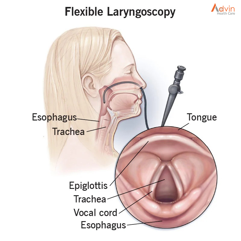
HIV-AIDS

Human immunodeficiency virus (HIV) is an infection that attacks the body’s immune system, specifically the white blood cells called CD4 cells. HIV destroys these CD4 cells, weakening a person’s immunity against opportunistic infections, such as tuberculosis and fungal infections, severe bacterial infections and some cancers.

WHO recommends that every person who may be at risk of HIV should access testing. People at increased risk of acquiring HIV should seek comprehensive and effective HIV prevention, testing and treatment services. HIV infection can be diagnosed using simple and affordable rapid diagnostic tests, as well as self-tests. It is important that HIV testing services follow the 5Cs: consent, confidentiality, counselling, correct results and connection with treatment and other services.

People diagnosed with HIV should be offered and linked to antiretroviral treatment (ART) as soon as possible following diagnosis and periodically monitored using clinical and laboratory parameters, including the test to measure virus in the blood (viral load). If ART is taken consistently, this treatment also prevents HIV transmission to others.

At diagnosis or soon after starting ART, a CD4 cell count should be checked to assess a person’s immune status. The CD4 cell count is a blood test used to assess progression of HIV disease, including risk for developing opportunistic infections and guides the use of preventive treatment. The normal range of CD4 count is from 500 to 1500 cells/mm3 of blood, and it progressively decreases over time in persons who are not receiving or not responding well to ART. If the person’s CD4 cell count falls below 200, their immunity is severely compromised, leaving them susceptible to infections and death. Someone with a CD4 count below 200 is described as having an advanced HIV disease (AHD).

HIV viral load measures the amount of virus in the blood. This test is used to monitor the level of viral replication and effectiveness of ART. The treatment goal is to reduce the viral load in the blood to undetectable levels (less than 50 copies/ml), and the persistent presence of detectable viral load (greater than 1000 copies/ml) in people living with HIV on ART is an indicator of inadequate treatment response and the need to change or adjust the treatment regimen.

The Spread of HIV


Transmission of HIV occurs through contact with certain body fluids of someone infected with HIV. These fluids include:

  • Blood
  • Semen
  • Vaginal fluid
  • Anal fluid
  • Breast milk

The most common form of HIV transmission in the United States is sharing drug injection equipment with or having sex with someone infected with HIV. Mothers with HIV can also pass the virus on to their child during pregnancy, childbirth and breastfeeding, but proper treatment greatly reduces this risk.

Prevention of HIV


Reduce your risk of becoming infected with HIV by practicing the following safe behaviors:

  • Correctly use condoms every time you have sex
  • Limit the number of sexual partners
  • Never share equipment for injecting drugs

Stages of HIV


There are three stages of HIV infection: acute HIV infection, clinical latency and AIDS. The infection gets worse as it progresses, eventually overwhelming your immune system without proper treatment. Progression through the stages occurs at different rates depending on a number of factors, including:

  • Genetic makeup
  • Level of health before infection
  • Amount of exposure to the virus
  • Genetic characteristics of the virus
  • How quickly treatment begins
  • Proper use of HIV medications
  • Health-related choices such as exercise, a healthy diet and not smoking

Stage 1: Acute HIV Infection

Two to four weeks after getting infected with HIV, people may develop severe flu-like symptoms. This is the acute HIV infection stage, which is also sometimes called “primary HIV infection” or “acute retroviral syndrome.” The symptoms can include:

  • Swollen glands
  • Fever
  • Rash
  • Sore throat
  • Joint and muscle aches and pains
  • Headache

Large amounts of HIV are being produced during this stage, so the number of CD4 cells drops rapidly. The immune response eventually brings the level of HIV down to a relatively stable level called the viral set point. Once the virus level drops, CD4 levels begin to rise, but usually don’t return to pre-infection levels. Due to the high levels of HIV, people in this stage are at a higher risk for transmitting the virus.

Stage 2: Clinical Latency

After the initial acute stage, HIV moves into the clinical latency stage, sometimes called “chronic HIV infection” or “asymptomatic HIV infection.” The term “latency” is used because the virus lives and reproduces at low levels within the infected individual without producing symptoms. Even though people are free of symptoms during the clinical latency stage, it’s still possible to transmit HIV to others; treatment helps reduce this risk.

Without treatment, the clinical latency stage usually lasts around 10 years, although people may progress through it faster or slower depending on a number of factors. When the viral load begins to rise again and CD4 levels fall, you eventually progress to the third and final stage of HIV infection.

Stage 3: AIDS

In the final stage of HIV infection, the immune system is badly damaged and the body becomes vulnerable to opportunistic infections. You have progressed to AIDS when at least one of the following occurs:

The level of CD4 cells falls to less than 200 cells per cubic millimeter of blood.

You develop one or more opportunistic infections.

People with AIDS usually survive about three years without treatment. Starting treatment after you have AIDS is helpful, but it’s more beneficial to begin treatment during one of the earlier stages. Most people with HIV in the United States rarely progress to AIDS thanks to effective treatment that controls the disease progression.

HIV Treatment


There is currently no cure for HIV, but people with HIV can live healthier, longer lives with proper treatment. Using HIV medicines to treat HIV is called antiretroviral therapy or ART. It includes a combination of HIV medicines taken every day. ART prevents the multiplication of HIV and lowers the amount of HIV in the body. Lower levels of HIV helps protect the immune system and keeps HIV infection from advancing to AIDS. Proper treatment also reduces the risk of transmitting HIV to others.

 Testing for HIV


The only way to determine whether or not you have HIV is to get tested. It’s important to know if you are infected because you can take steps to reduce the likelihood of transmitting HIV to others and start treatment. Early treatment is the best way to control the progression of HIV. If you notice flu-like symptoms after suspected exposure to HIV, then see a health care provider immediately. Because HIV infection can be difficult to detect with testing during the initial stage, it’s important to tell your healthcare provider that you think you’re at risk.

HIV/AIDS is a serious infection that attacks the immune system. However, with proper treatment, people infected with HIV can often live as long as someone without the infection.

Tobacco and Dental Health

The Effects of Tobacco Use on Oral Health


China consumes and produces more tobacco than any other country in the world. The next country in line is India. According to the WHO Global Report on Mortality, about 35 per cent of Indians over the age of 15 use tobacco and over 10 lakhs Indians die every year from tobacco-related diseases. Although the consumption of tobacco has declined over the past 2-3 years, the north-eastern states of India have been reported to have the highest consumption of tobacco in India.

Most of us know that tobacco consumption is harmful to health. A lot has been written about the effects of tobacco on health. Here we’ll talk about tobacco effects on our oral health. Some of the common oral health issues are as follows:

Bad Breath

The smoke particles left behind after smoking a cigarette stay in the mouth for quite some time leading to bad breath, which is commonly called stale smoker’s breath. Smoking or chewing tobacco also decreases the flow of saliva. Dry mouth is a breeding ground for bacteria thereby causing bad breath.

Tooth Discoloration

Nicotine is easily absorbed by your teeth. Although nicotine is colourless, it combines with oxygen to change the colour of the teeth from white to yellow. In people who chew tobacco, the nicotine combines with saliva to form a dark brown liquid which when allowed to stay in the mouth for long can stain the enamel.

Gum Disease


Smoking or chewing tobacco affects the attachment of the gums to the teeth. This is because the plaque formed near the gum line interferes with the gum tissue cells leaving your immune system compromised and making it harder for your body to fight off infections. The presence of nicotine reduces the blood flow to the gums, thus making you susceptible to oral infections. Particles left behind in the mouth increases the symptoms of dry mouth, providing an optimum environment for the bacteria to proliferate and cause inflammation. Tobacco use also affects the bone structure of the mouth as well as gum recession. These factors can increase your risk of jaw bone infection, which may lead to loss of teeth.

Lowered Success Rate of Dental Implants

A cosmetic procedure, such as dental implants, rely on having healthy, adequate bone in place to support the teeth. In smoker or tobacco users since the immune system is compromised the healing process take longer than usual. The same is the case if the tooth is extracted or if an oral surgery is performed. The recovery time is delayed.

Increased Chances of Developing Oral Cancer

Tobacco consumption contributes to 80 to 90% of diagnosed oral cancers. Therefore, mouth/jaw cancer is the number one reason why you should quit smoking or chewing tobacco. Although, tobacco causes many other health issues, they are less life threatening than cancer. Tobacco finds its way to the glands of your mouth and is easily filtered into your mouth tissues. Constant use of tobacco means constant absorption and filtration into the system making way for mouth cancer to spawn.

How to save yourself from facing these oral health issues? The answer is simple, just quit smoking or chewing tobacco. Here’s how you can overcome this habit:

Make a plan to quit the habit


You need to have a plan in place because with a plan handy it is easier to stay focused and motivated to quit. You can build your own plan or research online for a quit plan that’ll work for you.

Stay busy

Being busy is a great way to distract your mind and keep it occupied. Some of the activities you should try to keep you busy are:

  • Exercise
  • Movie or dinner with non-smoking friends and family
  • Walk
  • Chew gum
  • Take deep breaths
  • Drink lots of water
Stay away from triggers

People, things, places, and situations are your triggers that make you give in to your urge to consume tobacco. Here are some tips to stay away from the triggers:

  • Get rid of cigarettes, lighters, and ash trays or anything that remotely reminds you of tobacco.
  • Caffeine can make you jittery. Avoid it, try drinking water instead.
  • Rest well and eat healthy.
Stay positive

Quitting doesn’t happen in a day. The best approach to take is one day at a time. Most importantly, stay positive. Prepare your mind to quit first. Set a date and stick to it.

Ask for help from family and friends

Sometimes it gets difficult to rely solely on your willpower. Let your friends and family in. Ask them to support you on your plan to quit. Let them know what kind of support you’ll need from them. They can be a solid support, especially when you are going through a rough phase.

Prostate Cancer

Prostate cancer is a common type of cancer in males, but it is highly treatable in the early stages. It begins in the prostate gland, which sits between the penis and the bladder.

Prostate cancer is the most common cancer and the second leading cause of cancer death among men in the world. Prostate cancer usually grows very slowly, and finding and treating it before symptoms occur may not improve men’s health or help them live longer. Many prostate cancers grow slowly and are confined to the prostate gland, where they may not cause serious harm. However, while some types of prostate cancer grow slowly and may need minimal or even no treatment, other types are aggressive and can spread quickly.

Prostate cancer that’s detected early — when it’s still confined to the prostate gland — has the best chance for successful treatment.

What Is Prostate Cancer?


Cancer starts when cells in the body begin to grow out of control. Cells in nearly any part of the body can become cancer cells, and can then spread to other areas of the body.

Prostate cancer begins when cells in the prostate gland start to grow out of control. The prostate is a gland found only in males. It makes some of the fluid that is part of semen.

The prostate is below the bladder (the hollow organ where urine is stored) and in front of the rectum (the last part of the intestines). Just behind the prostate are glands called seminal vesicles that make most of the fluid for semen. The urethra, which is the tube that carries urine and semen out of the body through the penis, goes through the center of the prostate.

The size of the prostate can change as a man ages. In younger men, it is about the size of a walnut, but it can be much larger in older men.

Types of prostate cancer


Almost all prostate cancers are adenocarcinomas. These cancers develop from the gland cells (the cells that make the prostate fluid that is added to the semen).

Other types of cancer that can start in the prostate include:

  • Small cell carcinomas
  • Neuroendocrine tumors (other than small cell carcinomas)
  • Transitional cell carcinomas
  • Sarcomas

These other types of prostate cancer are rare. If you are told you have prostate cancer, it is almost certain to be an adenocarcinoma.

Some prostate cancers grow and spread quickly, but most grow slowly. In fact, autopsy studies show that many older men (and even some younger men) who died of other causes also had prostate cancer that never affected them during their lives. In many cases, neither they nor their doctors even knew they had it.

Possible pre-cancerous conditions of the prostate


Some research suggests that prostate cancer starts out as a pre-cancerous condition, although this is not yet known for sure. These conditions are sometimes found when a man has a prostate biopsy (removal of small pieces of the prostate to look for cancer).

Prostatic intraepithelial neoplasia (PIN)

In PIN, there are changes in how the prostate gland cells look when seen with a microscope, but the abnormal cells don’t look like they are growing into other parts of the prostate (like cancer cells would). Based on how abnormal the patterns of cells look, they are classified as:

  • Low-grade PIN: The patterns of prostate cells appear almost normal.
  • High-grade PIN: The patterns of cells look more abnormal.

Low-grade PIN is not thought to be related to a man’s risk of prostate cancer. On the other hand, high-grade PIN is thought to be a possible precursor to prostate cancer. If you have a prostate biopsy and high-grade PIN is found, there is a greater chance that you might develop prostate cancer over time.

PIN begins to appear in the prostates of some men as early as in their 20s. But many men with PIN will never develop prostate cancer.

Treatment


Treatment Trusted Source will depend on the cancer stage, among other factors, such as the Gleason score and PSA levels. It is also worth noting that many treatment options may be applicable, regardless of the stage of cancer.

In the sections below, we list some treatment options Trusted Source for prostate cancer and explore what treatment may mean for fertility.

Early-stage prostate cancer

If the cancer is small and localized, a doctor may recommend:

Watchful waiting or monitoring

The doctor may check PSA blood levels regularly but take no immediate action. Prostate cancer grows slowly, and the risk of treatment side effects may outweigh the need for immediate treatment.

Surgery

A surgeon may carry out a radical prostatectomy to remove the tumor. In addition to removing the prostate, the procedure may also involve the removal of the surrounding tissue, seminal vesicles, and nearby lymph nodes. A doctor can perform this procedure using either open, laparoscopic, or robot-assisted laparoscopic surgery.

Radiation therapy

This uses radiation to kill cancer cells or prevent them from growing. Options for early stage prostate cancer may include Trusted Source:

External radiation therapy: This method uses a machine outside the body to send radiation toward the cancer cells. Conformal radiation therapy is a type of external radiation that uses a computer to help guide and target a specific area, minimizing the risk to healthy tissue and allowing a high dose of radiation to reach the prostate tumor.

Internal radiation therapy: Also known as brachytherapy, this method uses radioactive seeds that a doctor implants near the prostate. A surgeon uses imaging scans, such as ultrasound or computed tomography to help guide the placement of the radioactive substance.

Treatment will depend on various factors. A doctor will discuss the best option for the individual.

Advanced prostate cancer

As cancer grows, it can spread throughout the body. If it spreads, or if it comes back after remission, treatment options will change. Options can include:

Chemotherapy: This option uses drugs to help stop the growth of cancer cells. While it can kill cancer cells around the body, it may cause Trusted Source adverse effects.

Hormonal therapy: Androgens are male hormones. The main androgens are testosterone and dihydrotestosterone. Blocking or reducing Trusted Source these hormones appears to stop or delay the growth of cancer cells. One option is to undergo surgery to remove the testicles, which produce most of the body’s hormones. Various drugs can also help.

Immunotherapy: This method uses a person’s immune system to help fight cancer. Scientists can use substances the body produces, or create them in a lab, to help boost or restore the body’s natural defenses against cancer.

Targeted therapy: This method uses drugs or other substances that identify and attack specific cancer cells.

Advin TURP Instrument Set


The Importance Of Pathology Laboratories

Pathology at a glance


Pathology connects science with medicine by studying the causes and progression of diseases. In addition, pathology laboratories assist doctors in researching and interpreting biopsy and other specimen results in a laboratory setting. Some pathology labs are standalone facilities, but they can also be found within hospitals to help a medical team understand better the causes of varied diseases and conditions.

The importance of clinical pathology


One of the major branches of this science is clinical pathology, while anatomic pathology is the other major branch. Doctors, scientists, and lab assistants working in pathology laboratories study samples from both branches to help medical professionals and doctors understand and control diseases better. Clinical pathology uses urine, blood, and other body fluid types samples. The doctors working in a clinical pathology lab use these samples for laboratory testing. They are used in blood banking tests, microbiology, immunology, clinical chemistry, and hematology tests.

How clinical pathology labs help doctors


These tests within the laboratory are essential because they help physicians understand better how a disease has progressed inside the human body. The tests performed in a clinical pathology lab help physicians understand and make good decisions about certain patients’ most appropriate and best treatments. Clinical pathology also helps physicians ensure they are making a correct diagnosis for a specific patient. Clinical Pathology techniques can be used to test whether a patient is suffering from a particular disease type and what treatment method will most effectively work in curing the disease. Clinical pathology takes the guesswork out of modern medicine.

Pathology laboratory final thoughts


Clinical pathology is an essential part of diagnosing and treating recommendations by your doctor. The treatment that your doctor recommends will be based on facts studied and interpreted through pathology test results to conclude the type of treatment method they should use in your care. Keep in mind that the experts, doctors, and other scientists working in pathology labs are experts on diseases. They have extensive knowledge about disease trends and are experts in preventing illnesses and diseases. Clinical Pathology is used to ensure that patients get the most accurate diagnosis for an illness or disease and receive the treatment they need. On top of it all, clinical pathology can also be an excellent prevention method for patients exploring ways to prevent disease occurrence, especially when they have a family history.

The value of clinical pathology labs and the doctors, scientists, and other staff who keep them operating effectively cannot be overstated. Their work helps diagnose, treat, and cure diseases that improve patients’ quality of life and save lives. This makes it vital for pathology labs to have state-of-the-art technology, optimal workflow, and safety measures in place to keep them operating at their highest potential.

Types of pathology labs


Hospital labs
Almost all hospitals contain a laboratory to support the clinical services offered at the hospital. The specific pathology services would include both anatomic (surgical pathology, cytopathology, autopsy) and clinical (laboratory medicine) pathology at most hospitals. Most, if not all, inpatient and many outpatients seen by hospital-affiliated physicians require tests performed by hospital labs.

Reference labs
Reference labs are usually private, commercial facilities that do both high volume and specialty (high complexity and/or rare) laboratory testing. Most of these tests are referred from physician’s offices, hospital facilities and other patient care facilities such as nursing homes. Reference labs, typically located at a site other than the healthcare facilities, are often used for specialized tests that are ordered only occasionally or require special equipment for analysis.

Public health labs
Public health laboratories are typically run by state and local health departments to diagnosis and protect the public from health threats such as outbreaks of infectious disease. These labs perform tests to monitor the prevalence of certain diseases in the community which are a public health concern, such as outbreaks of foodborne or waterborne illnesses or detection of unique infectious agents.

Maintaining Your Surgical Instruments

Proper care and handling are key to the longevity of your surgical instruments. After each procedure, you should be following a set cleaning process. Stainless steel instruments, and all other instruments, require proper care to maintain their properties.

Even though stainless-steel instruments are viewed as the best material, you can’t assume that they are entirely corrosion-resistant. The cleaning and sanitizing process of surgical instruments is complex and has become more difficult due to advancements; therefore, these instruments must be handled delicately.

Here are some tips to keep in mind when handling and cleaning your surgical instruments:

  • Immediately after use, clean and dry your instruments. Residue will cause staining if they are not rinsed right after the procedure. It is recommended that you use warm or cool, distilled water and solutions with a pH below 10 for cleaning, rinsing, and sterilization. Dry instruments thoroughly to minimize the risk of corrosion and water spots. For instruments such as forceps and scissors, make sure they are dried in an open position. Store instruments in dry areas.
  • Instruments should be used for their intended purpose only. Make sure you use the appropriate instruments for each procedure. Incorrect use can damage the instruments beyond repair or can impact its performance, which can lead to frustration and delays in the OR.
  • Don’t place instruments in saline or any other harsh solutions. Long-term exposure to saline can break down the surface of an instrument which can cause corrosion, thus, shortening its lifespan.
  • Use a softer brush for manual cleaning. Stiff plastic or nylon brushes are most effective for manual cleaning. You should pay special attention to any hard-to-reach areas and moving parts. Using a steel or wire brush will likely damage the instrument.
  • Lubricate all instruments that have any metal-to-metal action before autoclaving. Only use surgical instrument lubricates. Never use WD-40, oil, or other industrial lubricants. For instruments such as scissors or needle holders, sterilize them in an open position. Never lock an instrument during autoclaving – this will prevent the steam from reaching and sterilizing the metal-to-metal surfaces.
  • Ultrasonic cleaning is one of the most effective cleaning methods. Ultrasonic cleaning is the result of cavitation. The vibration waves create bubbles in the solution which grow until they finally implode, removing grime. Recommended exposure time is 5-10 minutes.

Importance Of CSSD In Hospitals

Each day, millions of medical procedures are executed in healthcare facilities around the world, with caregivers and patients relying on the availability and use of a wide range of supplies, instruments and equipment. These devices must be suitably cleaned, sanitized, and/or decontaminated, examined for quality to certify decent working conditions, and obtainable at the point of maintenance. In the absenteeism of appropriate management, processing and storage, these devices may become contaminated and minimize valuable patient care.

The Central Sterile Services Department (CSSD) is an organized place in hospitals and other health care services that performs sterilization and other activities on medical devices, equipment and consumables; for ensuing use by health workers in the operating theatre of the hospital and also for other aseptic processes, e.g., catheterization, wound stitching and bandaging in a medical, surgical, maternity or paediatric ward.

Sterilization is the procedure of destroying all living organisms on an article and is the chief duty of most sterile amenities departments. Objects to be sterilized must first be cleaned in an isolated decontamination room and inspected for efficiency, hygiene and damage. There are numerous procedures of sterilization, and which one is used is reliant on countless factors including: operational cost, latent hazards to workers, effectiveness, time, and arrangement of the articles being sterilized.

The CSSD has a significant part in patient care and in minimizing hospital surgical contamination. Various hospital departments depend on the service from the CSSD. With the centralization of the pre-disinfection, cleaning, packing and disinfection of all objects in one section, it is of supreme importance to deliver dependably high quality in the sterilization methods and product quality. As the quantity and variation of medical procedures and the kinds of medical devices are regularly mounting, improved processing is imperative for competence, economy and patient care.

Sterile processing departments are usually separated into 4 major areas to achieve the roles of sanitization, assemblage and sterile processing, disinfected storage, and dispersal.

Decontamination


  • Dismantlement & cleansing used surgical devices and other medical apparatus
  • Working and preserving special sanitization apparatus like automatic washers/purifiers, ultrasonic cleaners, etc.
  • Examining cleansed items to make sure they are hygienic
  • Assemblage
  • Operate PPE’s to shield self from getting harmful infections

Sterilization and Storage


  • Assemblage & packaging of cleaned and disinfected apparatuses
  • Disinfecting amassed trays of devices in suitable sterilizers
  • Precisely working and managing special sterilization equipment like autoclaves
  • Maintaining thorough records of figures of sterilized items, including Autoclave cycle statistics, lot/batch figures, expiration dates for forthcoming tracking of objects that have been sterilized, and stockpiled

Distribution


  • Providing crash carts
  • Maintaining decontaminated medical provisions
  • Confirming that disinfected goods do not become obsolete/averting incident linked sterility problems
  • Transporting sterile provisions where they are required and collecting contaminated ones
  • In most healthcare facilities, the central sterile supply department (CSSD) plays a crucial part in provisioning the items essential to provide quality patient care. To back infection control within the healthcare system, the CSSD staff members must be sufficiently trained and capable, and dedicated to doing the right thing. That means guaranteeing that shortcuts are never taken and that procedures and practices are dependably followed.
  • Applying advanced technologies and procedures may aid in increasing security for patients and staff. High barriers have to be passed in order to reproduce/imitate best systems known in the sterilization procedure. Structures should be in place to sustain and confirm that surgical instruments are appropriately managed before and after operation. Digital solutions for CSSD procedures permit for simplified data supervision and documentation. These keys may decrease mistakes, tension and pressure among the CSSD staff and simplify observance of procedures and strategies. In calculation, digital solutions aid instantaneous information updates and other well-organized behaviors to exchange information.
  • Centralizing the recycling of reusable apparatus helps safeguard undeviating ideals of practice, while also providing for enhanced workflow. This also simplifies the exercise and teaching of capable technicians who must be well-informed about the ethics, intricacies, trials, hazards, and procedures related with the CSSD function. Every CSSD task must be achieved in a way that shields the safety and wellbeing of patients, co-workers and the community.
  • Being employed in the sanitization department of CSSD necessitates detailed information and understanding of microbiology and the cleansing process; cleaning methods; phases in the cleaning procedure; aims of the cleaning method; levels of sanitization; documentation and cleaning of definite surgical instruments, syringes, needles, rubber objects, and specialty articles; cleaning substances and their use; sanitizing agents and their utilization; identification and effect of the cleaning process on diverse metals; different instrument lubrication and upkeep; appropriate clearance of all categories of waste; the conveyance of soiled items; and the operation of apparatus used in the cleaning procedure, such as washers, decontaminators, ultrasonic cleaners, cart washers, steam guns, scope washers, and so on.
  • The ideologies essential to attain sterilization must be understood and utilized. Sanitizers must be laden and worked properly, sterilization quality assurance procedures must be followed and understood to confirm that objects are sterile, and records must be preserved. Factors that can compromise sterile packaging must be understood, prohibited and punctually distinguished. Improving the quality administration with the latest advances helps to sustain a high standard in the CSSD, the “pillar” of sterile exercise in any hospital.

Advin CSSD Products


Pathology

Pathology is a branch of medical science primarily concerning the cause, origin, and nature of disease. It involves the examination of tissues, organs, bodily fluids, and autopsies in order to study and diagnose disease.

Currently, pathology can be divided into eight main areas, depending on the types of methods used or the types of diseases examined. These different disciplines are described below.

General pathology


General pathology describes a complex and broad field that involves the study of the mechanisms behind cell and tissue injury and understanding how the body responds to and repairs injury. Examples of areas that may be studied include necrosis, neoplasia, wound healing, inflammation and how cells adapt to injury. Thorough understanding in these areas is applied in the diagnosis of disease. General pathology is also the term used to describe anatomical and clinical pathology.

This field covers areas of pathology, but at a less specialist level. A person working in general pathology would be trained in the areas of laboratory analysis, such as hematology and clinical chemistry. However, they would have a less detailed knowledge than a person who specializes in one of these fields.

Anatomical pathology


This field is concerned with the study and diagnosis of illness through microscopic analysis of samples from bodily fluids, tissues organs and sometimes the entire body or autopsy. Factors that may be examined include the cell appearance, anatomical makeup and chemical signatures within cells.

This discipline can be subdivided into several disciplines and examples of these are given below:

  • Histology – Samples of bodily tissues and organs are prepared and examined in order to detect and diagnose disease. The architecture of tissue is observed at a microscopic level and the relationship between different cell and tissue types is examined.
  • Cytology – Bodily fluids and tissues are examined at the cellular level in order to screen for and diagnose disease and help aid treatment decisions. A cytologist will examine how cells look, form and function.
  • Forensic pathology – Forensic pathology is the examination of an autopsy in order to discover the cause of death. The external appearance is first assessed to check for evidence of wounds or suffocation, for example. Surgical procedures are then begun and the internal organs are studied to see whether internal injuries exist and are connected to external ones.

Clinical pathology


Also referred to as laboratory medicine, clinical pathology concerns the analysis of blood, urine and tissue samples to examine and diagnose disease. Examples of the information clinical pathology laboratories may provide include blood count, blood clotting and electrolyte results. A clinical pathologist is usually trained in microbiology, hematology or blood banking, but not at the same expert level as someone who specializes in one of these fields.

A clinical pathologist may come across problems that demand specific expertise, at which point they would need to consult a more specialized colleague. Clinical pathologists play a similar role to that of general pathologists, although they would not be involved in anatomical pathology.

Chemical Pathology or Biochemistry


Biochemists or chemical pathologists examine all aspects of disease, identifying changes in various different substances found in the blood and bodily fluids such as proteins, hormones and electrolytes since these changes can indicate and provide clues about disease or disease risk.

For example, a biochemist may assess cholesterol and triglyceride levels in order to determine heart disease risk. They may also look for and measure tumor markers, vitamins, poisons, medications and recreational drugs.

Genetics


There are three main branches of genetics and these include the following:

  • Cytogenetics: This is the analysis of chromosomal abnormalities at the microscopic level.
  • Biochemical genetics: The search for specific disease markers using biochemical techniques.
  • Molecular genetics: Gene mutations are searched for and analyzed using DNA technology.

Genetics involves performing tests on chromosomes, biochemical markers and DNA taken from bodily fluids and tissues in order to detect genetic illnesses.

Hematology


This field is concerned with various different disease aspects that affect the blood, including bleeding disorders, clotting problems, and anemia, for example. Another area of hematology is transfusion medicine, which involves performing blood typing, cross-matching for compatibility and managing large amounts of blood products. An example of a test a hematologist may perform is a blood clotting test to check whether a patient’s dose of warfarin needs increasing or decreasing.

Immunology


Immunologists perform immune function tests to establish whether or not a patient is suffering from an allergy and if so, what they are allergic to. Many diseases also arise as a result of the immune system having an abnormal reaction to healthy cells or tissues and launching an immune attack against them. This is referred to as a autoimmune disease. There is a range of immunological tests that can detect markers of autoimmune diseases such as rheumatoid arthritis, diabetes and lupus.

Microbiology


Microbiology is concerned with diseases caused by pathogenic agents such as bacteria, viruses, parasites and fungi. Samples of blood, bodily fluid and tissue are tested to establish whether infection exists, and the field of medical microbiology is also engaged with identifying new species of microorganisms.

Other areas encompassed by microbiology include control of infection outbreaks and researching the problems resulting from bacterial antibiotic resistance. One of the principal roles of the microbiologist is to make sure that antimicrobial drugs are prescribed and used appropriately.

Anaesthesia

Anesthesia is a treatment using drugs called anesthetics. These drugs keep you from feeling pain during medical procedures. Anesthesiologists are medical doctors who administer anesthesia and manage pain. Some anesthesia numbs a small area of the body. General anesthesia makes you unconscious (asleep) during invasive surgical procedures.

What is anesthesia?


Anesthesia is a medical treatment that keeps you from feeling pain during procedures or surgery. The medications used to block pain are called anesthetics. Different types of anesthesia work in different ways. Some anesthetic medications numb certain parts of the body, while other medications numb the brain, to induce a sleep through more invasive surgical procedures, like those within the head, chest, or abdomen.

How does anesthesia work?


Anesthesia temporarily blocks sensory/pain signals from nerves to the centers in the brain. Your peripheral nerves connect the spinal cord to the rest of your body.

Who performs anesthesia?


If you’re having a relatively simple procedure like a tooth extraction that requires numbing a small area, the person performing your procedure can administer the local anesthetic. For more complex and invasive procedures, your anesthetic will be administered by a physician anesthesiologist. This medical doctor manages your pain before, during and after surgery. In addition to your physician anesthesiologist, your anesthesia team can be comprised of physicians in training (fellows or residents), a certified registered nurse anesthetist (CRNA), or a certified anesthesiologist assistant (CAA).

What are the types of anesthesia?


The anesthesia your healthcare provider uses depends on the type and scope of the procedure. Options include:

  • Local anesthesia: This treatment numbs a small section of the body. Examples of procedures in which local anesthesia could be used include cataract surgery, a dental procedure or skin biopsy. You’re awake during the procedure.
  • Regional anesthesia: Regional anesthesia blocks pain in a larger part of your body, such as a limb or everything below your chest. You are can be conscious during the procedure, or have sedation in addition to the regional anesthetic. Examples include an epidural to ease the pain of childbirth or during a cesarean section (C-section), a spinal for hip or knee surgery, or an arm block for hand surgery.
  • General anesthesia: This treatment makes you unconscious and insensitive to pain or other stimuli. General anesthesia is used for more invasive surgical procedures, or procedures of the head, chest, or abdomen.
  • Sedation: Sedation relaxes you to the point where you will have a more natural sleep, but can be easily aroused or awakened. Light sedation can be prescribed by the person performing your procedure, or together with a regular nurse, if they both have training to provide moderate sedation. Examples of procedures performed with light or moderate sedation include cardiac catheterization and some colonoscopies. Deep sedation is provided by an anesthesia professional because your breathing may be affected with the stronger anesthetic medications, but you will be more asleep than with light or moderate sedation. Although you won’t be completely unconscious, you are not as likely to remember the procedure.

How should I prepare for anesthesia?


Make sure your healthcare provider has a current list of the medications and supplements (vitamins and herbal medications) you take. Certain drugs can interact with anesthesia or cause bleeding and increase the risk of complications. You should also:

  • Avoid food and drinks for eight hours before you go to the hospital unless directed otherwise.
  • Quit smoking, even if it’s just for one day before the procedure, to improve heart and lung health. The most beneficial effects are seen with no smoking for two weeks before.
  • Stop taking herbal supplements for one to two weeks before the procedure as directed by your provider.
  • Not take Viagra® or other medications for erectile dysfunction at least 24 hours before the procedure.
  • You should take certain (but not all) blood pressure medications with a sip of water as instructed by your healthcare provider.

What happens during anesthesia?


A physician anesthesiologist:

  • Administers one type or a combination of anesthetics listed above pain therapies, and possibly anti-nausea medications.
  • Monitors vital signs, including blood pressure, blood oxygen level, pulse and heart rate.
  • Identifies and manages problems, such as an allergic reaction or a change in vital signs.
  • Provides postsurgical pain management.

What should I do after getting anesthesia?


For procedures using local anesthesia, you can return to work or most activities after treatment unless your healthcare provider says otherwise. You’ll need more time to recover if you’ve received regional or general anesthesia or sedation. You should:

  • Have someone drive you home.
  • Rest for the remainder of the day.
  • Not drive or operate equipment for 24 hours.
  • Abstain from alcohol for 24 hours.
  • Only take medications or supplements approved by your provider.
  • Avoid making any important or legal decisions for 24 hours.

How long does it take to recover from anesthesia?


Anesthetic drugs can stay in your system for up to 24 hours. If you’ve had sedation or regional or general anesthesia, you shouldn’t return to work or drive until the drugs have left your body. After local anesthesia, you should be able to resume normal activities, as long as your healthcare provider says it’s okay.

Advin Anesthesia Product


Laryngoscopy

A laryngoscopy is an examination that gives your doctor a close-up view of your larynx and throat. The larynx is your voice box. It’s located at the top of your windpipe, or trachea.

It’s important to keep your larynx healthy because it contains your vocal folds, or cords. Air passing through your larynx and over the vocal folds causes them to vibrate and produce sound. This gives you the ability to speak.

A specialist known as an “ear, nose, and throat” (ENT) doctor will perform the exam. During the exam, your doctor place a small mirror into your throat, or insert a viewing instrument called a laryngoscope into your mouth. Sometimes, they’ll do both.

A laryngoscopy is a procedure healthcare providers use to examine your larynx (voice box). This is performed with a laryngoscope, a thin tube with a light, lens and a video camera that helps providers look closely at your larynx. Providers may do laryngoscopies in a clinic office or as surgery in an operating room.

What is my larynx?


Your larynx is located between your throat and your trachea (windpipe). Your larynx houses your vocal cords (vocal folds), which enable you to speak and sing. Your epiglottis sits on top of your larynx. Your epiglottis is a flap that closes when you eat or drink so items intended for your esophagus (food pipe) don’t end up in your larynx or in your airway. When people talk about something going down the wrong pipe, the pipe they’re talking about is your larynx.

When is laryngoscopy needed?


Your doctor may do it to find out why you have a sore throat that won’t go away or to diagnose an ongoing problem such as coughing, hoarseness, or bad breath. They also might do one when:

  • You have something stuck in your throat.
  • You have trouble breathing or swallowing.
  • You have an earache that won’t go away.
  • They need to examine something that could be a sign of a more serious health problem such as cancer.
  • They need to remove a growth.

Types of laryngoscopy


There are several ways your doctor may do this procedure:

Indirect laryngoscopy: This is the simplest form. Your doctor uses a small mirror and a light to look into your throat. The mirror is on a long handle, like the kind a dentist often uses, and it’s placed against the roof of your mouth.

The doctor shines a light into your mouth to see the image in the mirror. It can be done in a doctor’s office in just 5 to 10 minutes.

You’ll sit in a chair while the exam is done. Your doctor might spray something into your throat to make it numb. Having something stuck in your throat might make you gag, however.

Direct fiber-optic laryngoscopy: Many doctors now do this kind, sometimes called flexible laryngoscopy. They use a small telescope at the end of a cable, which goes up your nose and down into your throat.

It takes less than 10 minutes. You’ll get a numbing medication for your nose. Sometimes a decongestant is used to open your nasal passages as well. Gagging is a common reaction with this procedure as well.

Direct laryngoscopy: This is the most involved type. Your doctor uses a laryngoscope to push down your tongue and lift up the epiglottis. That’s the flap of cartilage that covers your windpipe. It opens during breathing and closes during swallowing.

Your doctor can do this to remove small growths or samples of tissue for testing. They can also use this procedure to insert a tube into the windpipe to help someone breathe during an emergency or in surgery.

Direct laryngoscopy can take up to 45 minutes. You’ll be given what’s called general anesthesia, so that you will not be awake during the procedure. Your doctor can take out any growths in your throat or take a sample of something that might need to be checked more closely.

I’m having a laryngoscopy what should I expect?


Your healthcare provider will consider your specific situation when deciding which type of laryngoscopy they’ll use. You may have your laryngoscopy in a clinic office or as a surgical procedure. For example, your provider may decide you should have a surgical laryngoscopy in an operating room. This is a direct laryngoscopy. Providers typically do direct laryngoscopies following in-office flexible laryngoscopies. Direct laryngoscopies may be done along with biopsies or other surgical procedures.

What happens before laryngoscopy?


If you’re having a surgical laryngoscopy, you’ll receive general anesthesia. Your provider will talk to you about getting ready for your procedure:

  • You may need to fast the night before your procedure.
  • You may need to avoid certain medications before your procedure.
  • If you smoke, your provider may recommend you stop smoking a week or so before your procedure.
  • You’ll need someone to give you a ride home after your surgery.

What happens during laryngoscopy?


Surgical laryngoscopies and clinic office laryngoscopies have different processes.

Laryngoscopy in an office clinic

  • In some cases, your provider may use a small, tilted mirror and a bright light to examine your vocal cords.
  • They also may use a flexible laryngoscope. This is a flexible fiberoptic scope inserted through your nose to look down your throat.
  • Your provider will apply a small amount of numbing medicine and decongestant to your nose. The numbing medication may taste bitter.
  • You may feel an urge to cough, but that urge is likely to subside.
  • Your provider will then gently pass the flexible laryngoscope into a nostril and look down your throat.
  • They may have you speak during the test to see how your voice box is working.
  • In some circumstances, they may use a special scope that’s inserted through your mouth.

Direct laryngoscopy in the operating room

Your provider will use a special laryngoscope that’s inserted through your mouth. Because you had general anesthesia, you won’t feel anything.

What happens after laryngoscopy?


Different things happen depending on whether you had a surgical laryngoscopy or one done in an office clinic.

In-office clinic laryngoscopy

  • Your nose and throat may feel numb for a few minutes after the procedure. This usually wears off in about 20 minutes.
  • You should avoid eating or drinking while your mouth and throat are numb.

Operating room direct laryngoscopy

  • You’ll go to the recovery unit after surgery to recover from anesthesia so your provider can watch for any complications or problems.
  • You may have a mild sore throat and/or hoarseness. Your provider may want you to rest your voice for a while after your surgery.

What are the benefits of a laryngoscopy?


Your doctor is concerned that you may have a problem in your larynx. A laryngoscopy is a good way of finding out if there is a problem.

If your surgeon finds a problem, they can perform biopsies (removing small pieces of tissue) to help make the diagnosis. For some people, the treatment can be performed at the same time.

For some people minor treatments can be performed at the same time.

Advin laryngoscopy products


Functional Endoscopic Sinus Surgery

Functional endoscopic sinus surgery (FESS) is a minimally invasive procedure which uses nasal endoscopes to enlarge the nasal drainage pathways of the paranasal sinuses to improve sinus ventilation and allow access of topical medications. This procedure is generally used to treat inflammatory and infectious sinus diseases, including chronic rhino sinusitis that do not respond to drugs, nasal polyps, some cancers, and decompression of eye sockets/optic nerve in Graves ophthalmopathy. In the surgery, an otolaryngologist removes the uncinate process of the ethmoid bone, while visualizing the nasal passage using a fiber optic endoscope. FESS can be performed under local anesthesia as an outpatient procedure. Generally patients experience only minimal discomfort during and after surgery. The procedure can take from 2 to 4 hours to complete.

Medical applications


Functional endoscopic sinus surgery is most commonly used to treat chronic rhinosinusitis (CRS), only after all non-surgical treatment options such as antibiotics, topical nasal corticosteroids, and nasal lavage with saline solutions[3] have been exhausted. (CRS) is an inflammatory condition in which the nose and at least one sinus become swollen and interfere with mucus drainage It can be caused by anatomical factors such as a deviated septum or nasal polyps (growths), as well as infection. Symptoms include difficulty breathing through the nose, swelling and pain around the nose and eyes, postnasal drainage down the throat, and difficulty sleeping. CRS is a common condition in children and young adults.

The purpose of FESS in treatment of CRS is to remove any anatomical obstructions that prevent proper mucosal drainage. A standard FESS includes removal of the uncinate process, and opening of the ethmoid air cells and Haller cells as well as the maxillary ostium, if necessary. If any nasal polyps obstructing ventilation or drainage are present, they are also removed. In the case of paranasal sinus/nasal cavity tumors (benign or cancerous), an otolaryngologist can perform FESS to remove the growths, sometimes with the help of a neurosurgeon, depending on the extent of the tumor. In some cases, a graft of bone or skin is placed by FESS to repair damages by the tumor.

In the thyroid disorder known as Graves’ ophthalmopathy, inflammation and fat accumulation in the orbitonasal region cause severe proptosis. In cases that have not responded to corticosteroid treatment, FESS can be used to decompress the orbital region by removing the ethmoid air cells and lamina papyracea. Bones of the orbital cavity or portions of the orbital floor may also be removed. endoscopic approach to FESS is a less invasive method than open sinus surgery, which allows patients to be more comfortable during and after the procedure. Entering the surgical field via the nose, rather than through an incision in the mouth as in the previous Caldwell-Luc method, decreases risk of damaging nerves which innervate the teeth. Because of its less-invasive nature, FESS is a common option for children with CRS or other sinonasal complications.

It has been suggested that one of the main objectives in FESS surgery is to allow for the introduction of local therapeutic agents (such as steroids) to the sinuses. Research has shown that a special modification of the nozzle of the nasal spray in patients who had FESS allows for better delivery of local therapeutic agents into the ethmoid sinuses.

What is the difference between ESS and FESS?


ESS or Endoscopic Sinus Surgery is a broader term which covers any endoscopic surgery of the nose and sinuses, including FESS. ESS may not be functional (e.g preservation of mucosa) and some indications for ESS include resection of sinonasal tumour, resection of skull base or brain tumour through the nose and sinuses, surgery of the eye through the nose and sinuses and surgery on the nasal septum.

Why do people call it functional endoscopic sinus surgery?


Functional endoscopic sinus surgery is also called endoscopic sinus surgery. Some healthcare providers use the term “functional” because the surgery is done to restore how your sinuses work, or function.

When would I need functional endoscopic sinus surgery?


Your healthcare provider may recommend FESS if you have chronic sinus inflammation or a chronic sinus infection that doesn’t improve with medical treatments, such as antibiotics and medications to manage allergies.

You develop sinusitis when the tissue that lines your sinuses begins to swell, trapping mucus that typically flows through your sinuses and out through your nose. The trapped fluid can grow bacteria that can cause infections. Healthcare providers may also recommend surgery if you have nasal polyps.

What happens before this procedure?


Your healthcare provider will let you know what to do before your surgery. Every person’s situation is different, but most healthcare providers recommend the following:

  • If you smoke, stop smoking at least three weeks before your surgery. Smoking can make your sinus symptoms worse. Ask your healthcare provider for advice or resources to help with this.
  • Don’t take aspirin for at least 10 days before your surgery. Even small amounts of aspirin can increase how much you bleed during and after your surgery.
  • If your surgery involves general anesthesia, don’t eat or drink anything after midnight the day of your surgery.
  • Your healthcare provider will administer general anesthesia just before your surgery begins.

How is functional endoscopic surgery performed?


FESS is the standard procedure to treat serious sinus conditions. Healthcare providers continue to refine their approach. Here’s an overview of the process:

  • Your healthcare provider puts decongestant medication in your nose.
  • They do a follow-up nasal endoscopy.
  • They inject a numbing solution into your nose.
  • Using the endoscope, they gently enter your nose. They insert surgical tools alongside the endoscope to use the endoscope to remove bone, diseased tissue or polyps that may be blocking your sinuses.
  • They may also use a small rotating burr to scrape out tissue.
  • Finally, your healthcare provider may pack your nose with material to absorb any blood or discharge.

What is the recovery after FESS?


After sinus surgery, the surgeon will usually place dressings in the sinus cavities to prevent post-operative adhesions and to minimise bleeding. These dressings may be absorbable (which do not require removal) or non-absorbable (which will usually be removed between 5 – 10 days after surgery). In some conditions, “nasal packs” may be inserted in the nasal cavities to minimise bleeding after surgery. These “nasal packs” are non absorbable and are usually removed in 1 – 2 days. Depending on the extent of the sinus operation, the patient may be allowed to go home on the same day (as a Day Surgery Procedure) or may be admitted to the ward and observed overnight. It is common for patients to feel blocked in the nose from the packing material and blood clots. A nasal gauze (“bolster”) will be placed under the nose to collect any blood that drips out (Fig 2). This is usually removed before the patient goes home. There may be mild pain after the surgery but this is usually well-controlled with pain relievers.

Advin FESS Instrument Product