Radiology Tests Explained

Radiology defines a wing of medicine that functions using radiant energy while diagnosing and treating diseases. In simple words, radiology means a process to determine whether a medical condition is present or not before moving ahead with the treatment. It is a test that takes images of different body parts and tries to find relevant issues to support a necessary diagnosis.

This branch of medicine comprises two areas – diagnostic radiology and interventional radiology. Both these forms use different radiant energy to provide a required diagnosis. Among the various radio imaging exams, some of the most common include- MRI, Ultrasound, X-ray, PET scan, etc. After the patients experience these tests, the radiologist will provide reports of their elucidations to the concerned doctors.

Now that individuals have a fair understanding of what radiology is, they should know about this diagnostic test’s different types and procedures.

The five most common radiological tests prescribed by doctors are explained below.

X-Rays


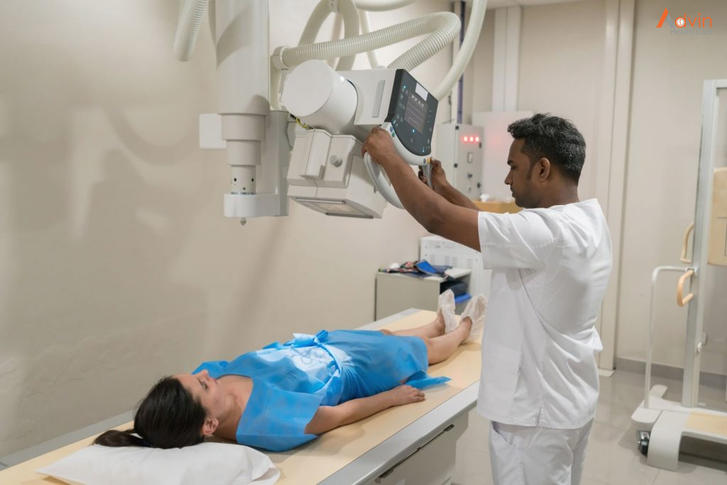
X-ray is the most common and widely performed radiological test. It is non-invasive and painless. This method makes use of electromagnetic radiation to detect even minor anomalies. Modern x-ray machines have become more efficient, with a ray source on one side and radiation-sensitive plates on the other. It helps in quick imaging and gives instant results.

The Test Procedure

The patient has to place the concerned body part between the ray source and the radiation plate. The doctor might ask them to move or turn to take images from different angles. This process takes only about 10 to 15 minutes.

Simple x-rays are done for screening as well as diagnostic purposes. It can:

  • detect hairline fractures, ligament tears, or lumps in certain areas
  • diagnose conditions like osteoporosis, arthritis, digestive tract issues, and infections
  • track the severity of an injury and the effects of treatment on the patient’s body
  • screening for and diagnosing the malignancy of breast lumps (mammogram)
  • live imaging of blood flow, hard and soft tissues, and joints using a contrast dye like barium (fluoroscopy/barium swallow radiology)

Ultrasound


Ultrasound, also called sonography, is another widespread radiological procedure. It uses high-frequency sound waves to detect different structures inside the body. There are no radiation waves in this test which makes this a safe procedure even for pregnant women. Ultrasound can be minimally invasive or non-invasive and mainly studies soft tissue structures and organs.

The Test Procedure

The doctor applies a gel on the target body part and places a transducer or probe which emits ultrasound waves. They move the probe around the body part to get a clear picture. For endoscopic ultrasound, the probe is inserted into the body through the patient’s mouth or rectum, depending on the part to be studied. The procedure can take about 25 minutes to 1 hour.

Purpose Of The Test

Ultrasound is used for diagnostic and interventional purposes. It is performed to:

  • diagnose breast lumps, joint inflammation, and issues related to gall bladder and prostate
  • diagnose inflammation or cancer in the digestive tract
  • study the progression of a pregnancy
  • act as a guide during biopsies

CT/CAT Scan


A computerised Axial Tomography scan uses a series of ionising radiations to form a cross-sectional image of the patient’s whole body. It is a non-invasive procedure and helps study almost all body structures.

The Test Procedure

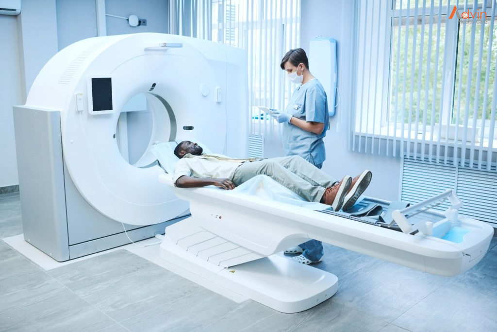
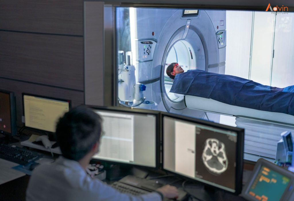
The patient is made to lie on a table that slides into the doughnut-shaped scanner. A scanning tube at the centre rotates around the patient’s body and creates a 3-D image. There is a transmitter inside the scanner through which the patient can talk to the doctor in case of nervousness or discomfort. The imaging takes only 15 to 20 minutes.

Purpose Of The Test

Doctors usually prescribe a CAT scan when other tests indicate some abnormality. It is done for diagnostic and radiology treatment purposes like:

  • studying the severity of a traumatic injury or fracture
  • detecting tumours and other abnormalities
  • diagnosing infections and heart and vascular diseases
  • guiding biopsies

MRI Scan


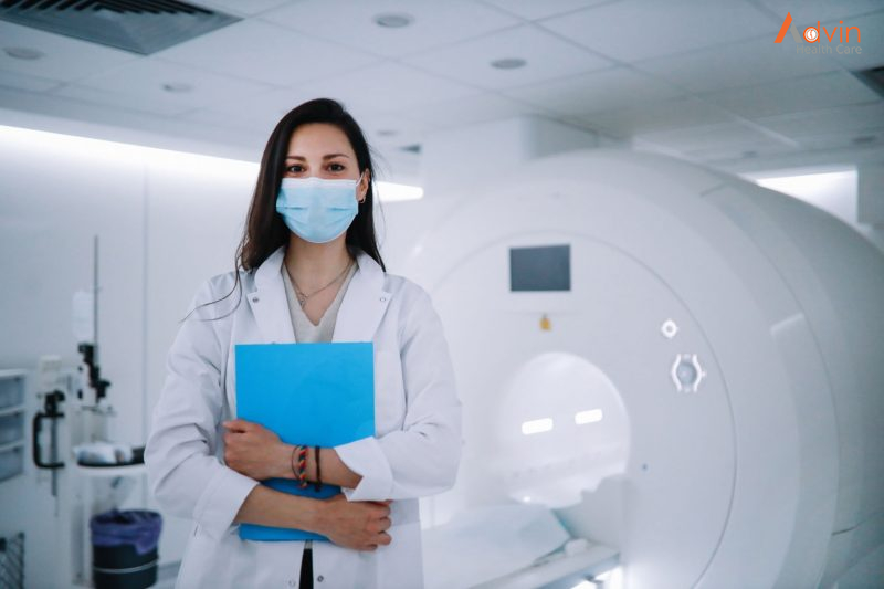
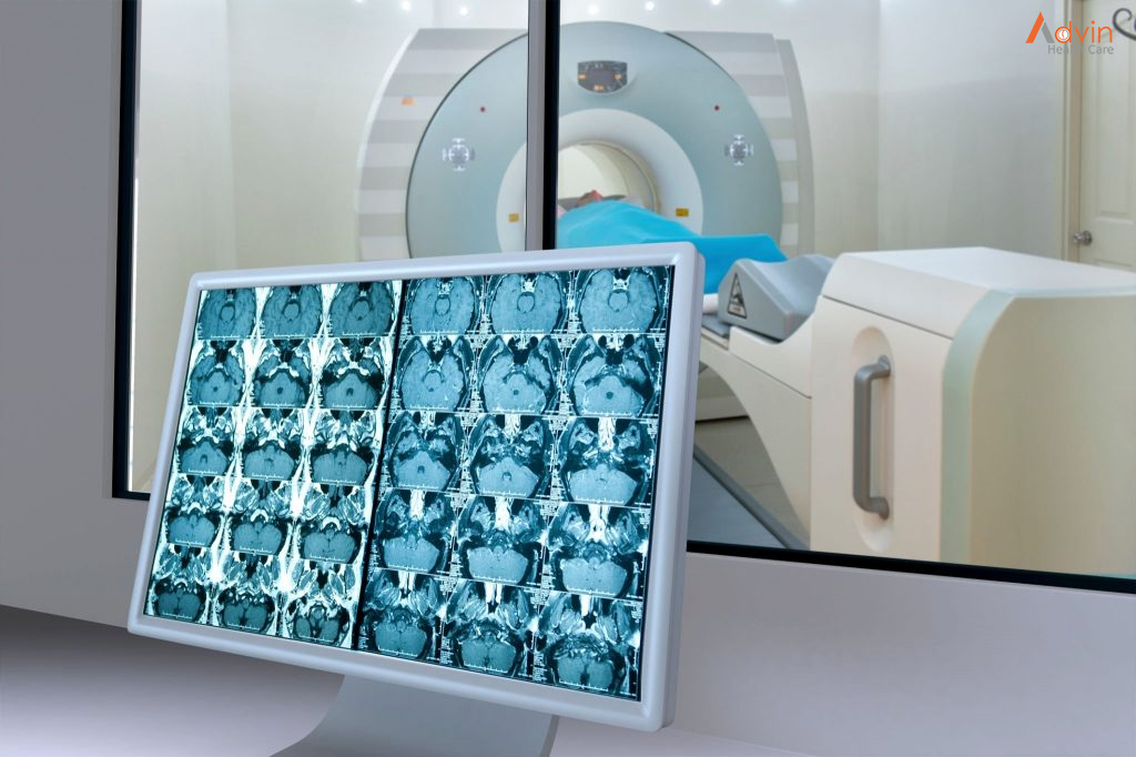
Magnetic Resonance Imaging works very similarly to a CAT scan. Only, it uses magnetic fields instead of ionising radiation. This test works for almost all body parts and conditions.  Due to the absence of radiation, it is also considered a safer method. However, it takes longer than a CAT scan.

The Test Procedure

The patient is made to lie on the sliding table, which moves into the MRI scanner. The scanner rotates, sending magnetic waves to create a 3-D image of the patient’s body. As it does so, the scanner makes loud noises. The patient can interact with the doctor through the transmitter in case of discomfort.

Purpose Of The Test

MRI scan helps in diagnosing a wide array of conditions. It includes:

  • spinal cord diseases
  • issues related to soft tissues like organs, blood vessels, joints, and tendons
  • strokes, aneurysms, and multiple sclerosis

PET Scan


Positron Emission Tomography scan follows a different methodology than the ones discussed above. In this test, a radioactive drug, called a radiotracer, is inserted into the patient’s body. The PET scanner studies the transmission of this tracer on a cellular level. This radiography test helps study any abnormalities and the functioning of different body systems.

The Test Procedure

The radiotracers are introduced into the patient’s body via an injection into the veins or inhaling gases, or drinking a mixture containing radiotracers. It depends on the organ system or body part to be studied. After about an hour of introducing the radiotracer into the body, the scanning is done. The whole procedure might take up to  2 hours. The radiotracers are introduced into the patient’s body via an injection into the veins, inhaling gases, or drinking a mixture containing radiotracers.

Purpose Of The Test

PET scan is a highly efficient procedure for performing diagnostic imaging. It is used to diagnose:

  • cancer
  • cardiovascular diseases
  • Alzheimer’s disease
  • Parkinson’s disease
  • digestive tract diseases
  • seizures
  • epilepsy

Future Technology in Orthopedic

Virtual reality, sensors and 3D printing for a better follow-up of the patient throughout his journey


If virtual reality can already be used to treat certain phobias, or even to help with rehabilitation thanks to mirror neurons, for example, the application of this technology combined with sensors and 3D printing could improve patient care throughout the course of their treatment, from diagnosis to post-operation follow-up.

Before the orthopedic operation and as a preventive measure, sensors could be used to monitor the patient’s physical data. For example, we could imagine a custom-made corset, which integrates sensors to follow in real time the evolution of the curvature of the back, as well as the use of post-operative implants capable of detecting possible complications.

Virtual reality technologies can also be used to plan and simulate the operation to be performed in a virtual environment, allowing the patient to experiment with several procedures in order to select the most appropriate one.

Virtual and mixed reality for the learning and training of doctors in orthopedics


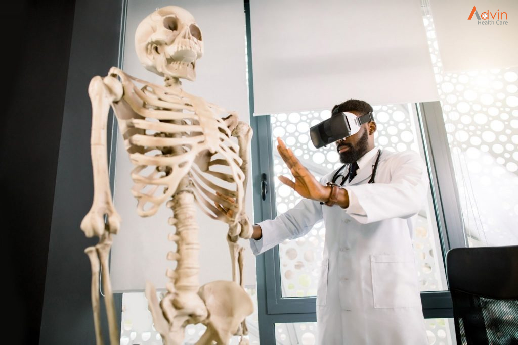
Originally, virtual reality in health was developed for educational purposes, for example by allowing a 3D reconstruction of the human body, to study in detail the different components and mechanisms of the body. This technology allows, for example, reconstruction by organ, and access to high quality educational information.

The development of virtual reality is revolutionizing surgical training methods: little by little, traditional methods such as teaching in the operating room or cadaver labs are being abandoned in favor of virtual reality, which provides a better viewpoint and a better understanding of the manipulations to be performed. This trend has become even more pronounced since the Covid-19 crisis, during which virtual reality has made it easier to train future surgeons.

Mixed reality, on the other hand, allows access to an immersive tutorial during an operation, to ensure that the correct actions are repeated.

Robotics and augmented mixed reality for greater precision in the orthopedic surgery operating room


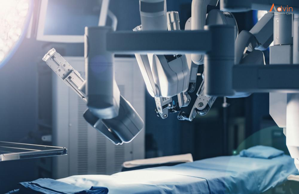
By introducing robotics, as well as augmented and virtual reality in the operating room, it is possible to significantly reduce variability during operations, to guarantee better results for patients.

The use of mixed reality in the operating room for orthopedic surgery, allowing the surgeon to be faster and more precise, is more recent. It was notably Prof. Thomas Gregory, in 2017, who demonstrated the interest of this technology in the operating room for orthopedics. Indeed, he operated on a shoulder prosthesis, assisted by mixed reality glasses, projecting a digital model on the patient, and gathering all the information collected during imaging examinations. The information available included, for example, the exact thickness of the tissue, or the precise location of surrounding organs invisible to the naked eye.

How Hospital Monitor Trolleys Useful?

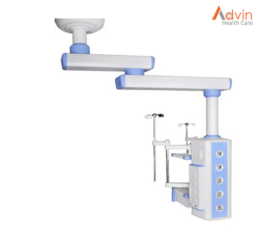
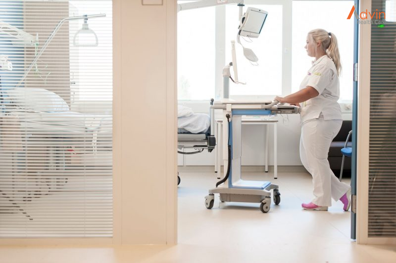
The hospital monitor trolley is considered to be one of the most useful and adaptable equipment. They play a variety of critical functions in providing quick and effective healthcare, and they are vital in numerous ways. Medical personnel across the globe are increasing their work efficiency by using them.

Hospital monitor trolleys are essential to medical institutions across the world. With their help, health facilities, treatment centres, and other medical institutions are able to provide high-quality care. Hospital monitor trolleys play a variety of roles that have an immediate positive influence on the effectiveness of care provided by hospitals.

The healthcare sector relies on a variety of high-end technologies to increase the workflow and efficiency of its medical staff. Stainless steel hospital monitor trolleys are commonly used in hospitals due to their high quality and resistance to corrosion, particularly in places where hygiene is of utmost priority. Though there are different kinds of trolleys available in the market, hospitals prefer the ones made of stainless steel because its strength and sterility makes these trolleys a centrepiece in medical technologies with clear advantages. Let’s take a look at those advantages in detail:.

Eases Cleaning and Disinfection

A dependable and sterile hospital monitor trolley does not jeopardise the hospital environ’s cleanliness and sanitation. The spills are simple to clean and does not invite germs, mildew, and rot. Healthcare facilities house a significant number of people suffering from transmissible diseases. So, the risk of transmission and infection is extremely high.

Top-notch hospital monitor trolleys made of stainless steel are electro-polished. So, they are almost always the preferred material for medical centres. Its key advantage is that it increases the trolley’s sanitary grade. It retains its lustre for long and its exterior holds contaminants at bay. It is scratch-resistant and easier to sterilise and cleanse. All of these features turn it into a great choice for medical centres.

Prevents Waste Reduction

By streamlining the procedure of arranging and accessing hospital instruments, high-quality hospital monitor trolleys assist hospitals in minimising surgical and clinical waste. As a result, it assists workers in keeping materials organised and easily accessible if needed.

Optimises Mobility & Portability

Hospital monitor trolleys also provide a secure and dependable means of moving multiple objects or equipment in one go. It is crucial to effectively move equipment properly since some of them may well be sharp and pose a safety concern to employees. Furthermore, because medical equipment are high-value objects, they must be carried in medical trolleys with a minimal chance of damage or substitution. Trolleys with wheels make it easier for employees to manoeuvre them across the halls with minimal effort.

Allows Carrying of Medicines & Equipment

In case you happen to be a healthcare professional who has to place orders, you may notice that a lot of the medication required by patients tends to weigh quite a lot. You wouldn’t want to have to carry a heavy load of medicine or lose time searching for hospital equipment while you’re in the midst of a patient’s care. At this juncture, a hospital monitor trolley will help you carry the medicine swiftly.If medical carts are built of steel alloys, they will last long. Stainless steel is perhaps the strongest material suitable for healthcare carts in the market.

Simplifies Storage & Increases Security

An added advantage of purchasing a hospital monitor trolley is that it may be used for storage. Employees in a healthcare facility need vital medications and supplies in hand. Trolleys are simple to set up and operate, and they provide a consistent experience for employees in healthcare. In addition to providing complete control over the trolley’s motions, the lockable structure can ensure protection while transporting medical supplies.

Promotes Diverse Range of Usages

Hospital monitor trolleys come in a variety of styles and sizes. This gives every medical institution the ability to acquire one that is appropriate for its need. Trolleys are versatile and sturdy, allowing them to be used in a variety of settings in the medical sector. For specific requirements or demands, the trolley must meet specified dimensions or standards.

Tips for Choosing the Best Hospital Monitor Trolley


Hospital monitor trolleys come in a variety of shapes and colours to meet the needs of the hospitals. They often have two or more shelves with wheels for ease of movement. As safety features, several of them have locking mechanisms. Here’s how you can choose the right one.

  • Take into account the portability of the trolley.
  • Purchase it from a well-known hospital furniture manufacturer in INDIA.
  • Consider the ease with which it can be assembled or disassembled.
  • Ensure the wheels and brakes are functioning properly.
  • Double-check for high-quality steel used in making the trolley.
  • Pay attention to the quality of the locks in it.
  • Choose a model with an ergonomic design for ease of sanitisation.

Advin Hospital Monitor Trolley


Diagnostic Kits

In vitro diagnostic tests have made it possible to accurately identify the specific microorganisms that cause disease and to perform additional testing to guide treatment selection. As such, diagnostic testing kits have become essential for diagnosing and monitoring disease, providing prognoses, and predicting treatment outcome. The global diagnostic market continues to grow, at a rate of 15% per year, and it forecasts to keep growing with the recent emergence of at-home diagnostic kits.

What Are Diagnostic Kits?


A diagnostic test is a medical test that is performed to diagnose or detect a specific disease or condition. This is in contrast to prognostic tests that predicts the likelihood of developing a disease or specific likely events happening during the course of that disease. These tests often come as pre-packaged test kits, which typically contain reagents and other items necessary to conduct the test to detect the presence of or to measure the levels of a given biomarker. These kits can also provide genetic information regarding mutations, deletions, and other abnormalities. Kits that test for genetic markers are often designed based on a newly discovered link between a patient genotype and the disease phenotype. Companies line Bio-Rad, Qiagen, and Abbott are some of the leaders in the diagnostic kit market.

Kits serve a variety of purposes and provide clinicians with a wide range of information, including the likelihood of a patient developing a complex disease over time, such as cancer, heart disease, and diabetes. In effect, these kits allow the physician to advise practical changes in lifestyle that may minimize future health risks or maximize preventative medical care. In addition to diagnosing disease, testing can also help determine the optimal course of treatment, specific to the patient in question. This includes more comprehensive diagnostic tests that can evaluate samples on the genomic level and can be used to analyze multiple genetic factors.

Diagnostic kits can serve various purposes


Diagnostic: These tests are used to confirm when a person has signs or symptoms of a genetic disease.

Predictive: A predictive test is used to search for genetic mutations linked with a specific condition, and indicates a person’s propensity to develop a disease before any symptoms are present. Moreover, complementary predictive diagnostic testing may provide information about how useful a therapeutic treatment may be in treating disease, but is not required to prescribe the drug, whereas companion testing is needed to prescribe it.

Pre-symptomatic: Pre-symptomatic tests are similar to predictive tests. These tests are used to determine risk for genetic conditions already known to be present in their family but show no symptoms.

Pharmacogenomic: Pharmacogenomic screening is a type of genetic test that may indicate a person’s response to certain types of drug treatment. This is still only available for a limited number of drugs, including Warfarin and Tamoxifen.

At-Home Diagnostic Kits


At-home diagnostic kits help people perform tests in the comfort of their house and typically provide rapid results, sometimes in as little as a minute. These kits can include health monitoring equipment that can be used to monitor blood pressure and insulin levels in diabetic patients. The increased adoption of these self-help and do-it-yourself kits is due to convenience and the ability to obtain results quickly, at an affordable cost. While doubts among end-users remain about their overall reliability, rapid testing kits, such as those for COVID-19, are increasingly being used by the public. As such, the at-home testing kit market is expected to reach $8M by 2028, with high competition in the market driving the increase.

Medical device companies continue to develop new diagnostic kits for use in the lab and at home, serving a wide range of populations. With the growing geriatric population, rising awareness of personal health, and an increased interest in human genetics, the demand for diagnostic kits is projected to grow in the coming years, driven in part by higher demand for at-home diagnostic tools. As exemplified by the demand for rapid COVID-19 testing kits, the call for at-home diagnostic tools is likely to remain high; however, the current lack of reimbursement policies and high production costs will likely create roadblocks in meeting the short- and long-term demand for these diagnostics devices.

Rules & Regulation OF Diagnostic Lab


Laboratory developed tests (LDTs) are not commercially marketed to other entities or sold as medical devices in the diagnostics market. Though the FDA has stated they believe LDTs to be “medical devices,” the laboratories that develop them argue they are more aptly described as “testing services” and should not fall under the FDA’s regulation. The FDA typically does not actively enforce most of its regulations on laboratories that offer these services. However, they do impose certain restrictions over the sale of specific reagents used in the kits to these labs. Under these regulations, the reagent manufacturers must meet the regulations rather than the laboratories themselves.

Conversely, genetic in vitro diagnostic kits are commercially marketed by medical device manufacturers as testing kits sold to laboratories, which perform the tests. They can include reagents, collection devices, and measurement apparatuses, and fall under either Class II or Class III “medical devices” of the FDA’s Federal Food, Drug, and Cosmetic Act. While Class II in vitro kits must satisfy slightly less stringent requirements, Class II devices face stringent requirements set by the FDA, including approval before sale.

In vitro kits must also meet specific labeling requirements. This includes clear and concise labeling of the immediate container as well as any inserts and the outer packaging. The container should identify the product, lot number, intended use, a statement of warning where applicable, and for reagents, concentration, storage, and manipulation instructions, as well as a means to assure that the product meets appropriate standards of purity, such as an expiration date. The outer package label should include the established product name, a summary or explanation of the test, and identify whether it’s a qualitative or quantitative test.

Smart Hospital

With digital technology becoming the norm of daily life, it’s not surprising to see it make its way into the healthcare industry. Digital technology is being used in hospitals to help patients interact with doctors. Many hospitals have already implemented electronic health records, so any healthcare professional can access the information anywhere at any time, such as the hospital, doctor’s offices, patients’ homes, etc.

What Is A Smart Hospital?


There are three crucial layers that will help improve the quality of care – data, insight and access security entities. A smart hospital would make it easier to interact with patients, but the challenge to bringing this technology “to life” is how we can implement into existing hospitals. Because the infrastructure is aging, and IT systems are piecemealed over a number of years, making integration difficult.

A smart hospital is one equipped with a digital brain (artificial intelligence) that meets the needs of all the patients. Movies such as IRobot, Resident Evil and others touch on smart buildings. But instead of focusing on the “evil” aspects of these buildings, focus on what a smart hospital could do.

For example, the building recognizes you have a broken leg and sets up appointments with the right medical team and helps with the readying of the equipment, medical supplies and treatment room. From there, it tells you when your appointment is with the staff.

If a smart hospital is to be developed, the building must be changed to handle the Internet of Things (IoT) so that physical objects can be combined with digital technology. A central digital platform is the only way all these can be successfully combined to create a smart hospital.

This is attainable through a human-developed design based on works in hospitals and what could be better. Looking at various hospital surveys, people who feel like they’re heard or get a response to their concerns are much happier about the experience.

Developing a digital strategy that combines an array of building, medical and operational systems while also allowing patient access to much-needed information on various devices isn’t easy.

Benefits Of A Smart Hospital Management System


Improve patient satisfaction.

Mobile applications from a bedside tablet can be tied into the building management system to give patients control over their own room temperature and lighting, as well as the ability to call the nurse, view noise levels, and control their smart TV.

Improved Diagnosis and Treatment

The primary benefit of a smart hospital management system is that it improves patient diagnosis and treatment. The patient’s health report and medical history from the past to the present, as well as the illness he is suffering from and the care he received, can all be added to and accessed via the HMS. Using this knowledge, doctors can better assess patients’ health problems, ultimately empowering them to provide the best care possible.

Availability of Information At Fingertips

The hospital’s data enables you to track and monitor various events to improve your approach and set performance targets. With a smart hospital management system, you will have all of the details you need at your fingertips, including the patient’s medical history, inventory, finance, workforce, and more. You can use this knowledge to figure out what flaws there are in the system or in which department and how to fix them. The information also aids you in developing a long-term strategy for your healthcare facility.

Reduce Unnecessary Expenses

When all of the hospital’s operations are handled manually, there is a high risk of information leakage and irregularities. Because, as humans, we are prone to making mistakes. These errors will have serious consequences in the long run. However, with the aid of a smart hospital management system, you can easily control all of the hospital’s operations, even with fewer human resources. Moreover, this will also limit human interference in the system, resulting in lower operational costs and less leakage.

Enhance productivity.

A digital infrastructure utilizes forward thinking network connectivity to enhance wireless communication and transfer of digital data such as electronic medical records and digital imaging. Being able to access patient information remotely through smartphones and tablets enables hospital staff to react efficiently and quickly, improving not on productivity, but also patient care.

Protect them like they are your own.

With a digital, intelligent infrastructure, hospital security teams can integrate video surveillance, access control, intercom, intruder detection, fire safety, RTLS, among other security systems to provide real-time data and alarms. In addition, in the event a security incident occurs, actionable reports with traceability are available for forensic analysis.

Increased Data Protection

Another significant advantage of hospital management systems is improved data protection. Since everything is done through a secure system, only authorized individuals have access to the specific data collection. Furthermore, in a cloud-based hospital management system, everything is interconnected, ensuring that there are no risks of data loss and that the patients’ medical history or condition information remains completely protected.

Real-time Data Access

Since a smart hospital management system is a centralized system, doctors, administrative personnel, and other employees can access data in real-time, enabling them to make the best decisions possible. For example, the real-time data about the inventory will assist management in replenishing and updating the inventory. Similarly, the real-time data about the patient’s treatment will assist doctors in making clinical decisions with no room for error or ambiguity.

HIV-AIDS

Human immunodeficiency virus (HIV) is an infection that attacks the body’s immune system, specifically the white blood cells called CD4 cells. HIV destroys these CD4 cells, weakening a person’s immunity against opportunistic infections, such as tuberculosis and fungal infections, severe bacterial infections and some cancers.

WHO recommends that every person who may be at risk of HIV should access testing. People at increased risk of acquiring HIV should seek comprehensive and effective HIV prevention, testing and treatment services. HIV infection can be diagnosed using simple and affordable rapid diagnostic tests, as well as self-tests. It is important that HIV testing services follow the 5Cs: consent, confidentiality, counselling, correct results and connection with treatment and other services.

People diagnosed with HIV should be offered and linked to antiretroviral treatment (ART) as soon as possible following diagnosis and periodically monitored using clinical and laboratory parameters, including the test to measure virus in the blood (viral load). If ART is taken consistently, this treatment also prevents HIV transmission to others.

At diagnosis or soon after starting ART, a CD4 cell count should be checked to assess a person’s immune status. The CD4 cell count is a blood test used to assess progression of HIV disease, including risk for developing opportunistic infections and guides the use of preventive treatment. The normal range of CD4 count is from 500 to 1500 cells/mm3 of blood, and it progressively decreases over time in persons who are not receiving or not responding well to ART. If the person’s CD4 cell count falls below 200, their immunity is severely compromised, leaving them susceptible to infections and death. Someone with a CD4 count below 200 is described as having an advanced HIV disease (AHD).

HIV viral load measures the amount of virus in the blood. This test is used to monitor the level of viral replication and effectiveness of ART. The treatment goal is to reduce the viral load in the blood to undetectable levels (less than 50 copies/ml), and the persistent presence of detectable viral load (greater than 1000 copies/ml) in people living with HIV on ART is an indicator of inadequate treatment response and the need to change or adjust the treatment regimen.

The Spread of HIV


Transmission of HIV occurs through contact with certain body fluids of someone infected with HIV. These fluids include:

  • Blood
  • Semen
  • Vaginal fluid
  • Anal fluid
  • Breast milk

The most common form of HIV transmission in the United States is sharing drug injection equipment with or having sex with someone infected with HIV. Mothers with HIV can also pass the virus on to their child during pregnancy, childbirth and breastfeeding, but proper treatment greatly reduces this risk.

Prevention of HIV


Reduce your risk of becoming infected with HIV by practicing the following safe behaviors:

  • Correctly use condoms every time you have sex
  • Limit the number of sexual partners
  • Never share equipment for injecting drugs

Stages of HIV


There are three stages of HIV infection: acute HIV infection, clinical latency and AIDS. The infection gets worse as it progresses, eventually overwhelming your immune system without proper treatment. Progression through the stages occurs at different rates depending on a number of factors, including:

  • Genetic makeup
  • Level of health before infection
  • Amount of exposure to the virus
  • Genetic characteristics of the virus
  • How quickly treatment begins
  • Proper use of HIV medications
  • Health-related choices such as exercise, a healthy diet and not smoking

Stage 1: Acute HIV Infection

Two to four weeks after getting infected with HIV, people may develop severe flu-like symptoms. This is the acute HIV infection stage, which is also sometimes called “primary HIV infection” or “acute retroviral syndrome.” The symptoms can include:

  • Swollen glands
  • Fever
  • Rash
  • Sore throat
  • Joint and muscle aches and pains
  • Headache

Large amounts of HIV are being produced during this stage, so the number of CD4 cells drops rapidly. The immune response eventually brings the level of HIV down to a relatively stable level called the viral set point. Once the virus level drops, CD4 levels begin to rise, but usually don’t return to pre-infection levels. Due to the high levels of HIV, people in this stage are at a higher risk for transmitting the virus.

Stage 2: Clinical Latency

After the initial acute stage, HIV moves into the clinical latency stage, sometimes called “chronic HIV infection” or “asymptomatic HIV infection.” The term “latency” is used because the virus lives and reproduces at low levels within the infected individual without producing symptoms. Even though people are free of symptoms during the clinical latency stage, it’s still possible to transmit HIV to others; treatment helps reduce this risk.

Without treatment, the clinical latency stage usually lasts around 10 years, although people may progress through it faster or slower depending on a number of factors. When the viral load begins to rise again and CD4 levels fall, you eventually progress to the third and final stage of HIV infection.

Stage 3: AIDS

In the final stage of HIV infection, the immune system is badly damaged and the body becomes vulnerable to opportunistic infections. You have progressed to AIDS when at least one of the following occurs:

The level of CD4 cells falls to less than 200 cells per cubic millimeter of blood.

You develop one or more opportunistic infections.

People with AIDS usually survive about three years without treatment. Starting treatment after you have AIDS is helpful, but it’s more beneficial to begin treatment during one of the earlier stages. Most people with HIV in the United States rarely progress to AIDS thanks to effective treatment that controls the disease progression.

HIV Treatment


There is currently no cure for HIV, but people with HIV can live healthier, longer lives with proper treatment. Using HIV medicines to treat HIV is called antiretroviral therapy or ART. It includes a combination of HIV medicines taken every day. ART prevents the multiplication of HIV and lowers the amount of HIV in the body. Lower levels of HIV helps protect the immune system and keeps HIV infection from advancing to AIDS. Proper treatment also reduces the risk of transmitting HIV to others.

 Testing for HIV


The only way to determine whether or not you have HIV is to get tested. It’s important to know if you are infected because you can take steps to reduce the likelihood of transmitting HIV to others and start treatment. Early treatment is the best way to control the progression of HIV. If you notice flu-like symptoms after suspected exposure to HIV, then see a health care provider immediately. Because HIV infection can be difficult to detect with testing during the initial stage, it’s important to tell your healthcare provider that you think you’re at risk.

HIV/AIDS is a serious infection that attacks the immune system. However, with proper treatment, people infected with HIV can often live as long as someone without the infection.

Tobacco and Dental Health

The Effects of Tobacco Use on Oral Health


China consumes and produces more tobacco than any other country in the world. The next country in line is India. According to the WHO Global Report on Mortality, about 35 per cent of Indians over the age of 15 use tobacco and over 10 lakhs Indians die every year from tobacco-related diseases. Although the consumption of tobacco has declined over the past 2-3 years, the north-eastern states of India have been reported to have the highest consumption of tobacco in India.

Most of us know that tobacco consumption is harmful to health. A lot has been written about the effects of tobacco on health. Here we’ll talk about tobacco effects on our oral health. Some of the common oral health issues are as follows:

Bad Breath

The smoke particles left behind after smoking a cigarette stay in the mouth for quite some time leading to bad breath, which is commonly called stale smoker’s breath. Smoking or chewing tobacco also decreases the flow of saliva. Dry mouth is a breeding ground for bacteria thereby causing bad breath.

Tooth Discoloration

Nicotine is easily absorbed by your teeth. Although nicotine is colourless, it combines with oxygen to change the colour of the teeth from white to yellow. In people who chew tobacco, the nicotine combines with saliva to form a dark brown liquid which when allowed to stay in the mouth for long can stain the enamel.

Gum Disease


Smoking or chewing tobacco affects the attachment of the gums to the teeth. This is because the plaque formed near the gum line interferes with the gum tissue cells leaving your immune system compromised and making it harder for your body to fight off infections. The presence of nicotine reduces the blood flow to the gums, thus making you susceptible to oral infections. Particles left behind in the mouth increases the symptoms of dry mouth, providing an optimum environment for the bacteria to proliferate and cause inflammation. Tobacco use also affects the bone structure of the mouth as well as gum recession. These factors can increase your risk of jaw bone infection, which may lead to loss of teeth.

Lowered Success Rate of Dental Implants

A cosmetic procedure, such as dental implants, rely on having healthy, adequate bone in place to support the teeth. In smoker or tobacco users since the immune system is compromised the healing process take longer than usual. The same is the case if the tooth is extracted or if an oral surgery is performed. The recovery time is delayed.

Increased Chances of Developing Oral Cancer

Tobacco consumption contributes to 80 to 90% of diagnosed oral cancers. Therefore, mouth/jaw cancer is the number one reason why you should quit smoking or chewing tobacco. Although, tobacco causes many other health issues, they are less life threatening than cancer. Tobacco finds its way to the glands of your mouth and is easily filtered into your mouth tissues. Constant use of tobacco means constant absorption and filtration into the system making way for mouth cancer to spawn.

How to save yourself from facing these oral health issues? The answer is simple, just quit smoking or chewing tobacco. Here’s how you can overcome this habit:

Make a plan to quit the habit


You need to have a plan in place because with a plan handy it is easier to stay focused and motivated to quit. You can build your own plan or research online for a quit plan that’ll work for you.

Stay busy

Being busy is a great way to distract your mind and keep it occupied. Some of the activities you should try to keep you busy are:

  • Exercise
  • Movie or dinner with non-smoking friends and family
  • Walk
  • Chew gum
  • Take deep breaths
  • Drink lots of water
Stay away from triggers

People, things, places, and situations are your triggers that make you give in to your urge to consume tobacco. Here are some tips to stay away from the triggers:

  • Get rid of cigarettes, lighters, and ash trays or anything that remotely reminds you of tobacco.
  • Caffeine can make you jittery. Avoid it, try drinking water instead.
  • Rest well and eat healthy.
Stay positive

Quitting doesn’t happen in a day. The best approach to take is one day at a time. Most importantly, stay positive. Prepare your mind to quit first. Set a date and stick to it.

Ask for help from family and friends

Sometimes it gets difficult to rely solely on your willpower. Let your friends and family in. Ask them to support you on your plan to quit. Let them know what kind of support you’ll need from them. They can be a solid support, especially when you are going through a rough phase.

Prostate Cancer

Prostate cancer is a common type of cancer in males, but it is highly treatable in the early stages. It begins in the prostate gland, which sits between the penis and the bladder.

Prostate cancer is the most common cancer and the second leading cause of cancer death among men in the world. Prostate cancer usually grows very slowly, and finding and treating it before symptoms occur may not improve men’s health or help them live longer. Many prostate cancers grow slowly and are confined to the prostate gland, where they may not cause serious harm. However, while some types of prostate cancer grow slowly and may need minimal or even no treatment, other types are aggressive and can spread quickly.

Prostate cancer that’s detected early — when it’s still confined to the prostate gland — has the best chance for successful treatment.

What Is Prostate Cancer?


Cancer starts when cells in the body begin to grow out of control. Cells in nearly any part of the body can become cancer cells, and can then spread to other areas of the body.

Prostate cancer begins when cells in the prostate gland start to grow out of control. The prostate is a gland found only in males. It makes some of the fluid that is part of semen.

The prostate is below the bladder (the hollow organ where urine is stored) and in front of the rectum (the last part of the intestines). Just behind the prostate are glands called seminal vesicles that make most of the fluid for semen. The urethra, which is the tube that carries urine and semen out of the body through the penis, goes through the center of the prostate.

The size of the prostate can change as a man ages. In younger men, it is about the size of a walnut, but it can be much larger in older men.

Types of prostate cancer


Almost all prostate cancers are adenocarcinomas. These cancers develop from the gland cells (the cells that make the prostate fluid that is added to the semen).

Other types of cancer that can start in the prostate include:

  • Small cell carcinomas
  • Neuroendocrine tumors (other than small cell carcinomas)
  • Transitional cell carcinomas
  • Sarcomas

These other types of prostate cancer are rare. If you are told you have prostate cancer, it is almost certain to be an adenocarcinoma.

Some prostate cancers grow and spread quickly, but most grow slowly. In fact, autopsy studies show that many older men (and even some younger men) who died of other causes also had prostate cancer that never affected them during their lives. In many cases, neither they nor their doctors even knew they had it.

Possible pre-cancerous conditions of the prostate


Some research suggests that prostate cancer starts out as a pre-cancerous condition, although this is not yet known for sure. These conditions are sometimes found when a man has a prostate biopsy (removal of small pieces of the prostate to look for cancer).

Prostatic intraepithelial neoplasia (PIN)

In PIN, there are changes in how the prostate gland cells look when seen with a microscope, but the abnormal cells don’t look like they are growing into other parts of the prostate (like cancer cells would). Based on how abnormal the patterns of cells look, they are classified as:

  • Low-grade PIN: The patterns of prostate cells appear almost normal.
  • High-grade PIN: The patterns of cells look more abnormal.

Low-grade PIN is not thought to be related to a man’s risk of prostate cancer. On the other hand, high-grade PIN is thought to be a possible precursor to prostate cancer. If you have a prostate biopsy and high-grade PIN is found, there is a greater chance that you might develop prostate cancer over time.

PIN begins to appear in the prostates of some men as early as in their 20s. But many men with PIN will never develop prostate cancer.

Treatment


Treatment Trusted Source will depend on the cancer stage, among other factors, such as the Gleason score and PSA levels. It is also worth noting that many treatment options may be applicable, regardless of the stage of cancer.

In the sections below, we list some treatment options Trusted Source for prostate cancer and explore what treatment may mean for fertility.

Early-stage prostate cancer

If the cancer is small and localized, a doctor may recommend:

Watchful waiting or monitoring

The doctor may check PSA blood levels regularly but take no immediate action. Prostate cancer grows slowly, and the risk of treatment side effects may outweigh the need for immediate treatment.

Surgery

A surgeon may carry out a radical prostatectomy to remove the tumor. In addition to removing the prostate, the procedure may also involve the removal of the surrounding tissue, seminal vesicles, and nearby lymph nodes. A doctor can perform this procedure using either open, laparoscopic, or robot-assisted laparoscopic surgery.

Radiation therapy

This uses radiation to kill cancer cells or prevent them from growing. Options for early stage prostate cancer may include Trusted Source:

External radiation therapy: This method uses a machine outside the body to send radiation toward the cancer cells. Conformal radiation therapy is a type of external radiation that uses a computer to help guide and target a specific area, minimizing the risk to healthy tissue and allowing a high dose of radiation to reach the prostate tumor.

Internal radiation therapy: Also known as brachytherapy, this method uses radioactive seeds that a doctor implants near the prostate. A surgeon uses imaging scans, such as ultrasound or computed tomography to help guide the placement of the radioactive substance.

Treatment will depend on various factors. A doctor will discuss the best option for the individual.

Advanced prostate cancer

As cancer grows, it can spread throughout the body. If it spreads, or if it comes back after remission, treatment options will change. Options can include:

Chemotherapy: This option uses drugs to help stop the growth of cancer cells. While it can kill cancer cells around the body, it may cause Trusted Source adverse effects.

Hormonal therapy: Androgens are male hormones. The main androgens are testosterone and dihydrotestosterone. Blocking or reducing Trusted Source these hormones appears to stop or delay the growth of cancer cells. One option is to undergo surgery to remove the testicles, which produce most of the body’s hormones. Various drugs can also help.

Immunotherapy: This method uses a person’s immune system to help fight cancer. Scientists can use substances the body produces, or create them in a lab, to help boost or restore the body’s natural defenses against cancer.

Targeted therapy: This method uses drugs or other substances that identify and attack specific cancer cells.

Advin TURP Instrument Set


The Importance Of Pathology Laboratories

Pathology at a glance


Pathology connects science with medicine by studying the causes and progression of diseases. In addition, pathology laboratories assist doctors in researching and interpreting biopsy and other specimen results in a laboratory setting. Some pathology labs are standalone facilities, but they can also be found within hospitals to help a medical team understand better the causes of varied diseases and conditions.

The importance of clinical pathology


One of the major branches of this science is clinical pathology, while anatomic pathology is the other major branch. Doctors, scientists, and lab assistants working in pathology laboratories study samples from both branches to help medical professionals and doctors understand and control diseases better. Clinical pathology uses urine, blood, and other body fluid types samples. The doctors working in a clinical pathology lab use these samples for laboratory testing. They are used in blood banking tests, microbiology, immunology, clinical chemistry, and hematology tests.

How clinical pathology labs help doctors


These tests within the laboratory are essential because they help physicians understand better how a disease has progressed inside the human body. The tests performed in a clinical pathology lab help physicians understand and make good decisions about certain patients’ most appropriate and best treatments. Clinical pathology also helps physicians ensure they are making a correct diagnosis for a specific patient. Clinical Pathology techniques can be used to test whether a patient is suffering from a particular disease type and what treatment method will most effectively work in curing the disease. Clinical pathology takes the guesswork out of modern medicine.

Pathology laboratory final thoughts


Clinical pathology is an essential part of diagnosing and treating recommendations by your doctor. The treatment that your doctor recommends will be based on facts studied and interpreted through pathology test results to conclude the type of treatment method they should use in your care. Keep in mind that the experts, doctors, and other scientists working in pathology labs are experts on diseases. They have extensive knowledge about disease trends and are experts in preventing illnesses and diseases. Clinical Pathology is used to ensure that patients get the most accurate diagnosis for an illness or disease and receive the treatment they need. On top of it all, clinical pathology can also be an excellent prevention method for patients exploring ways to prevent disease occurrence, especially when they have a family history.

The value of clinical pathology labs and the doctors, scientists, and other staff who keep them operating effectively cannot be overstated. Their work helps diagnose, treat, and cure diseases that improve patients’ quality of life and save lives. This makes it vital for pathology labs to have state-of-the-art technology, optimal workflow, and safety measures in place to keep them operating at their highest potential.

Types of pathology labs


Hospital labs
Almost all hospitals contain a laboratory to support the clinical services offered at the hospital. The specific pathology services would include both anatomic (surgical pathology, cytopathology, autopsy) and clinical (laboratory medicine) pathology at most hospitals. Most, if not all, inpatient and many outpatients seen by hospital-affiliated physicians require tests performed by hospital labs.

Reference labs
Reference labs are usually private, commercial facilities that do both high volume and specialty (high complexity and/or rare) laboratory testing. Most of these tests are referred from physician’s offices, hospital facilities and other patient care facilities such as nursing homes. Reference labs, typically located at a site other than the healthcare facilities, are often used for specialized tests that are ordered only occasionally or require special equipment for analysis.

Public health labs
Public health laboratories are typically run by state and local health departments to diagnosis and protect the public from health threats such as outbreaks of infectious disease. These labs perform tests to monitor the prevalence of certain diseases in the community which are a public health concern, such as outbreaks of foodborne or waterborne illnesses or detection of unique infectious agents.

Maintaining Your Surgical Instruments

Proper care and handling are key to the longevity of your surgical instruments. After each procedure, you should be following a set cleaning process. Stainless steel instruments, and all other instruments, require proper care to maintain their properties.

Even though stainless-steel instruments are viewed as the best material, you can’t assume that they are entirely corrosion-resistant. The cleaning and sanitizing process of surgical instruments is complex and has become more difficult due to advancements; therefore, these instruments must be handled delicately.

Here are some tips to keep in mind when handling and cleaning your surgical instruments:

  • Immediately after use, clean and dry your instruments. Residue will cause staining if they are not rinsed right after the procedure. It is recommended that you use warm or cool, distilled water and solutions with a pH below 10 for cleaning, rinsing, and sterilization. Dry instruments thoroughly to minimize the risk of corrosion and water spots. For instruments such as forceps and scissors, make sure they are dried in an open position. Store instruments in dry areas.
  • Instruments should be used for their intended purpose only. Make sure you use the appropriate instruments for each procedure. Incorrect use can damage the instruments beyond repair or can impact its performance, which can lead to frustration and delays in the OR.
  • Don’t place instruments in saline or any other harsh solutions. Long-term exposure to saline can break down the surface of an instrument which can cause corrosion, thus, shortening its lifespan.
  • Use a softer brush for manual cleaning. Stiff plastic or nylon brushes are most effective for manual cleaning. You should pay special attention to any hard-to-reach areas and moving parts. Using a steel or wire brush will likely damage the instrument.
  • Lubricate all instruments that have any metal-to-metal action before autoclaving. Only use surgical instrument lubricates. Never use WD-40, oil, or other industrial lubricants. For instruments such as scissors or needle holders, sterilize them in an open position. Never lock an instrument during autoclaving – this will prevent the steam from reaching and sterilizing the metal-to-metal surfaces.
  • Ultrasonic cleaning is one of the most effective cleaning methods. Ultrasonic cleaning is the result of cavitation. The vibration waves create bubbles in the solution which grow until they finally implode, removing grime. Recommended exposure time is 5-10 minutes.