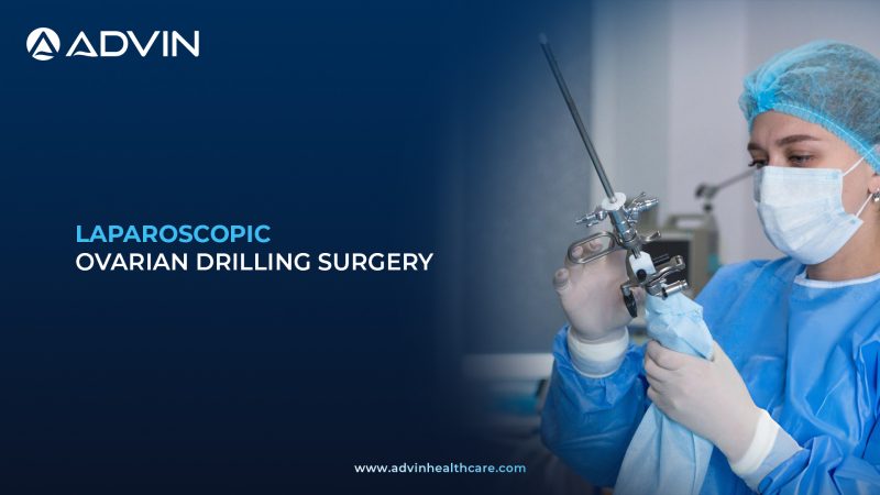
Overview of Laparoscopic Ovarian Drilling Procedure
Laparoscopic ovarian drilling is a minimally invasive surgery used to treat polycystic ovary syndrome (PCOS). It helps restore ovulation in women who do not respond to medication. This procedure can improve fertility and hormonal balance.
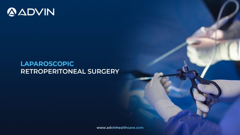
Minimally Invasive Laparoscopic Technique for PCOS Management
Laparoscopic ovarian drilling is performed under general anesthesia to ensure patient comfort and safety. Small incisions are made in the abdomen to insert a laparoscopic camera and surgical instruments. The camera provides a clear, magnified view of the ovaries. A fine needle electrode or laser is used to make small controlled punctures on the ovarian surface. These punctures reduce androgen-producing tissue and help restore ovulation. The procedure is completed with minimal trauma and short operating time.
Clinical Indications for Laparoscopic Ovarian Drilling
- To induce ovulation in women with PCOS resistant to medication
- To improve chances of natural conception
- To reduce excess androgen hormone production
- To regulate menstrual cycles in selected patients
Laparoscopic Instruments and Energy Devices for Ovarian Drilling
- Laparoscopic Camera System
- Laparoscopic Trocars
- Laparoscopic Graspers
- Needle Electrode or Laser Probe
- Electrosurgical Generator or Laser System
- Suction Irrigation System
- Laparoscopic Needle Holder
- Energy Sealing Device
- Uterine Manipulator
- Surgical Drapes
Leading Global Markets for Laparoscopic Ovarian Drilling
- India
- United States
- United Kingdom
- Germany
- Japan
Clinical and Commercial Advantages of Laparoscopic Ovarian Drilling
- Improves ovulation in medication-resistant PCOS
- Enhances natural fertility potential
- Reduces need for long-term hormonal treatment
- Minimally invasive with quick recovery
- Can restore more regular menstrual cycles
Advin Health Care is a globally leading manufacturer of laparoscopic ovarian drilling related products, providing reliable and high-quality solutions for advanced minimally invasive gynecological procedures.
Get Connected:
+91-70717 27261 | urology@advinhealthcare.com | www.advinhealthcare.com