Nitinol Guidewire vs Stainless Steel Guidewire: Which One Should You Choose?

Introduction

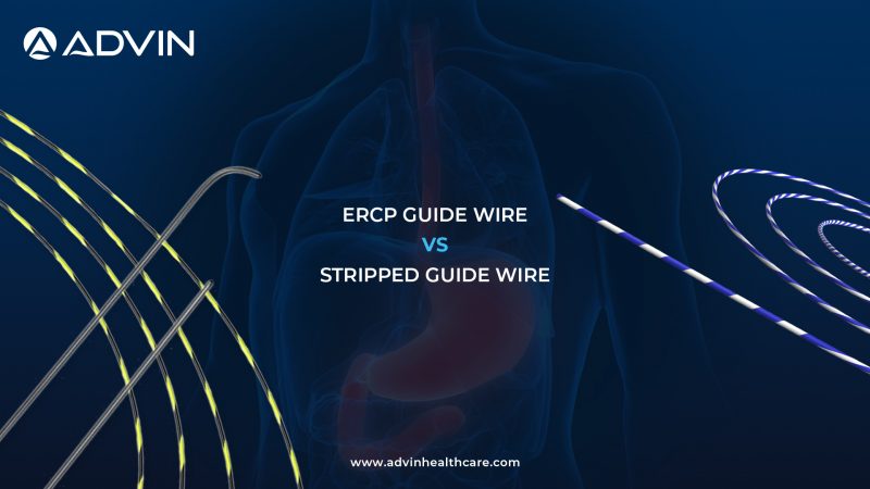
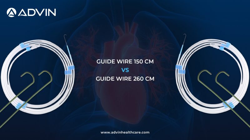
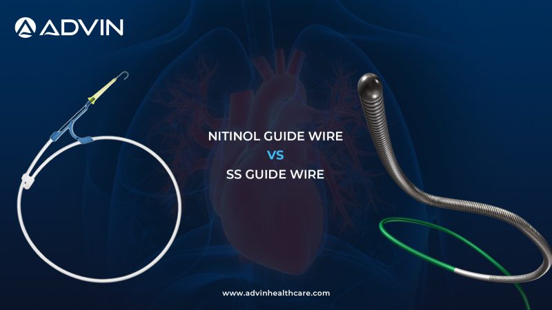
Guidewires are essential tools in interventional cardiology, providing support, navigation, and access within blood vessels. Among the most commonly used types are Nitinol Guidewires and Stainless Steel (SS) Guidewires.

While both serve the same purpose, their material properties significantly influence performance, flexibility, and control. Understanding these differences helps clinicians choose the right guidewire for specific procedural needs.

Product Overview

Nitinol Guidewire

Nitinol Guidewires are made from a nickel-titanium alloy known for its flexibility and shape memory. These guidewires are ideal for navigating complex and tortuous vessels with ease.

Stainless Steel (SS) Guidewire

Stainless Steel Guidewires are known for their strength and support. They provide excellent pushability and control, making them suitable for procedures requiring high precision and stability.

Quick Comparison Table

Feature Nitinol Guidewire SS Guidewire
Material Nickel-Titanium Alloy Stainless Steel
Flexibility High Moderate
Support Moderate High
Navigation Smooth in complex paths Precise control
Clinical Use Complex vessels Stable procedures

Key Feature Comparison

Nitinol Guidewire Features

  • High flexibility for navigating tortuous anatomy 
  • Shape memory property for maintaining structure 
  • Kink-resistant performance 
  • Smooth tracking through vessels 
  • Reduced risk of vessel trauma 
  • Ideal for complex and distal access 

Stainless Steel Guidewire Features

  • High strength and strong support 
  • Excellent pushability and torque control 
  • Provides stable device guidance 
  • Suitable for precise and controlled procedures 
  • Maintains rigidity during interventions 
  • Ideal for straightforward vascular paths 

Common Features

  • Provide reliable vascular access and navigation 
  • Support interventional cardiology procedures 
  • Compatible with standard devices and techniques 
  • Ensure smooth device delivery 
  • Designed for safe and efficient performance 
  • Sterile and single-use for patient safety 

Key Differences

  • Material: Nitinol vs Stainless Steel 
  • Flexibility: High vs Moderate 
  • Support: Moderate vs High 
  • Navigation: Smooth vs Controlled 
  • Use Case: Complex anatomy vs Stable procedures 

Which One Should You Choose?

Choose Nitinol Guidewire when

  • Navigating complex or tortuous vessels 
  • Flexibility and smooth tracking are required 
  • Reduced vessel trauma is important 

Choose Stainless Steel Guidewire when

  • Strong support and control are needed 
  • Procedures require precise navigation 
  • Stable and straightforward vessel access is involved 

Conclusion

Both Nitinol and Stainless Steel Guidewires play a critical role in interventional procedures. Nitinol offers flexibility and adaptability, while stainless steel provides strength and precision.

Selecting the right guidewire based on procedural needs ensures better control, improved outcomes, and enhanced patient safety.

Explore More Products>>

Get Connected:

+91-75037 27248 | cardiology@advinhealthcare.com | www.advinhealthcare.com

Laparoscopic Colectomy Set: Complete Solution for Minimally Invasive Colorectal Surgery

Clinical Purpose

The Laparoscopic Colectomy Set is a comprehensive minimally invasive surgical solution designed for the resection of the colon in cases of colorectal cancer, inflammatory bowel disease, diverticular disease, and other colonic pathologies. It integrates advanced laparoscopic visualization systems, electrosurgical and vessel sealing technologies, insufflation systems, precision instruments, and essential disposables to ensure safe dissection, effective vascular control, and efficient bowel resection with improved patient outcomes.

Evolution of Colorectal Surgery

Colorectal surgery has evolved from traditional open procedures to advanced laparoscopic techniques. Open colectomy required large incisions, resulting in increased pain, longer hospital stays, and higher complication rates.

With laparoscopic advancements, surgeons can now perform precise bowel resections using HD visualization and advanced energy devices. This evolution has significantly improved surgical accuracy, reduced blood loss, minimized post-operative complications, and enabled faster recovery with better cosmetic results.

Set Components and Functional Integration

Equipment

  • Advin Electrosurgical Unit 400 W – Advin Electro+
  • Advin Vessel Sealing System – Advin SafeSeal+
  • Advin CO2 Insufflator – Advin CO2+
  • Advin Full HD Camera System
  • Medical Monitor 24″ / 27″
  • LED Light Source
  • Fiber Optic Cable

Instruments

  • Laparoscope 10 mm / 30°
  • Veress Needle 2.1 mm
  • Trocar (All Types)
  • Laparoscopic Grasper
  • Laparoscopic Needle Holder
  • Laparoscopic Bipolar Forceps
  • Monopolar Electrodes (L Hook / Spatula)
  • Suction Irrigation Cannula Set
  • Laparoscopic Knot Pusher
  • Vessel Sealer with Cutter / Golden Tip

Disposables

  • Hem-O-Lock Clips
  • Titanium Clips
  • Laparoscopy Endo Bag
  • CO2 Insufflator Tubing
  • Connectors

Drapes

  • Laparoscopy Drape
  • Major Surgery Drape
  • Universal Split Drape

Clinical Applications

  • Colorectal cancer surgery
  • Colectomy (partial or total colon removal)
  • Diverticulitis and diverticular disease
  • Inflammatory bowel disease (Crohn’s disease, ulcerative colitis)
  • Bowel obstruction and benign colonic conditions

Instructions for Use (Laparoscopic Colorectal Surgery – Colectomy, Short & Smart Workflow)

  • Prepare the patient using laparoscopy, major surgery, and split drapes to maintain a sterile field. 
  • Set up the Advin CO2+ insufflator and create pneumoperitoneum using the Veress needle
  • Insert laparoscopic trocars to establish working ports. 
  • Connect the Advin Full HD Camera System, medical monitor, LED light source, and fiber optic cable for clear visualization. 
  • Introduce the 10 mm / 30° laparoscope and inspect the abdominal cavity. 
  • Use laparoscopic graspers to mobilize and expose the colon segment. 
  • Perform dissection using monopolar electrodes and bipolar forceps, supported by Advin Electro+
  • Seal and divide vessels using the Advin SafeSeal+ vessel sealer with cutter (Golden Tip)
  • Apply Hem-O-Lock or titanium clips where required for secure ligation. 
  • Resect the diseased colon segment and manage bowel ends as per surgical plan. 
  • Use the suction irrigation cannula to maintain a clean operative field. 
  • Place the specimen in an endo bag and remove it safely. 
  • Complete anastomosis using needle holder and knot pusher if required. 
  • Ensure hemostasis, release CO2, remove trocars, and close port sites securely. 
  • After completion, clean and sterilize reusable instruments and dispose of consumables properly.

Countries with High Clinical Adoption

  • United States
  • Germany
  • Japan
  • India
  • United Kingdom

Key Clinical Advantages

  • Minimally invasive colorectal surgery
  • Reduced blood loss and post-operative pain
  • Faster recovery and shorter hospital stay
  • Enhanced precision with HD visualization
  • Effective vascular control with advanced energy devices
  • Improved cosmetic outcomes

Advin Health Care Product Overview

Advin Health Care offers a complete Laparoscopic Colorectal Surgery Set designed for precision, safety, and efficiency in advanced gastrointestinal surgery. With high-performance electrosurgical systems, vessel sealing technology, reliable insufflation systems, and high-quality laparoscopic instruments, the set enables surgeons to perform safe and effective colectomy procedures while maintaining international quality standards.

Explore More Products>>

Get Connected:

+91-70717 27261 | urology@advinhealthcare.com | www.advinhealthcare.com

URS Alligator Forceps vs URS Triprong Forceps: Choosing the Right Instrument for Foreign Body and Stone Retrieval

Introduction: Grasping Mechanism Defines Retrieval Efficiency

In endourology and endoscopic procedures, forceps are essential for grasping, holding, and retrieving foreign bodies or stones. The choice between Alligator Forceps and Triprong Forceps significantly affects grip strength, precision, and retrieval success.

While both instruments serve similar purposes, they differ in jaw design, holding mechanism, and clinical application.

Product Overview in Clinical Use

Alligator Forceps

Alligator Forceps are designed with long, narrow jaws that open vertically, similar to an alligator’s mouth.

Key characteristics:

  • Serrated jaws 
  • Long, slim shaft 
  • Controlled opening mechanism 

Clinical advantages:

  • Strong and secure grip 
  • Precise handling in narrow spaces 
  • Suitable for linear or elongated objects 

They are commonly used in foreign body retrieval and stone handling in confined areas.

Triprong Forceps

Triprong Forceps feature three-pronged jaws that open outward to grasp objects from multiple sides.

Key characteristics:

  • Three flexible prongs 
  • Wide opening capability 
  • Multi-point contact 

Clinical advantages:

  • Better grip on irregular or round objects 
  • Increased surface contact 
  • Effective for loose or mobile stones 

They are widely used in stone retrieval and grasping objects with uneven shapes.

Workflow-Based Usage: Firm Grip vs Multi-Point Capture

Both forceps are used for retrieval but differ in gripping style:

  • Alligator Forceps → Linear, firm grip 
  • Triprong Forceps → Multi-directional grip 

This impacts:

  • Type of object handled 
  • Stability during extraction 
  • Ease of capture 

Core Differences That Drive Clinical Choice

The key difference lies in jaw design and gripping mechanism:

  • Alligator forceps offer precision and strong linear grip 
  • Triprong forceps provide multi-point grasping and better hold on irregular objects 

Quick Comparison Overview

Aspect Alligator Forceps Triprong Forceps
Jaw Design Two serrated jaws Three prongs
Grip Type Linear grip Multi-point grip
Precision High Moderate
Holding Strength Strong Moderate to strong
Object Type Linear / firm objects Round / irregular objects
Maneuverability High in narrow spaces Moderate
Clinical Use Foreign body retrieval Stone / fragment retrieval

Clinical Preference & Real-World Application

Alligator Forceps are commonly used in:

  • Foreign body removal 
  • Narrow anatomical pathways 
  • Situations requiring precise grip 

Their design ensures secure and controlled handling.

Triprong Forceps are preferred in:

  • Stone retrieval procedures 
  • Handling round or irregular objects 
  • Cases requiring multi-point grasp 

Their structure ensures better capture and reduced slippage.

Practical Selection Guide

  • Choose Alligator Forceps for precision and firm grip 
  • Choose Triprong Forceps for irregular objects and better capture 
  • Use alligator forceps in narrow spaces 
  • Use triprong forceps for stones and loose fragments 

Conclusion: Precision vs Versatility in Gripping

Both instruments are essential in endoscopic retrieval:

  • Alligator → Precise, strong, and controlled 
  • Triprong → Versatile, adaptive, and effective for irregular shapes 

The ideal choice depends on:

  • Type of object 
  • Anatomical location 
  • Required grip style 

Why Advin Health Care is the Right Choice

Advin Health Care offers a complete range of retrieval forceps designed for precision and reliability.

  • Alligator forceps provide strong grip and accurate handling 
  • Triprong forceps deliver versatile grasping and efficient retrieval 

With Advin, healthcare providers benefit from:

  • High-quality materials 
  • Durable construction 
  • Consistent clinical performance

Explore More Products>>

Get Connected:

+91-70717 27261 | urology@advinhealthcare.com | www.advinhealthcare.com

ERCP Guidewire vs Stripped Guidewire: Smooth Access vs Controlled Exchange in Biliary Procedures

Introduction: Why Guidewire Selection Impacts ERCP Success

Guidewires are the backbone of ERCP procedures, enabling access, navigation, and device placement within the biliary or pancreatic ducts. While both ERCP Guidewire and Stripped Guidewire are used in the same procedure, their roles differ based on the stage of intervention. During ERCP, a guidewire is first used to access the duct and then to support device exchange such as sphincterotomes, dilators, or stents. Choosing the right type at the right time improves control and procedural efficiency. This blog explains how each guidewire contributes to a smooth workflow.

Device Role Across Procedure Stages

ERCP Guidewire (Hydrophilic Guidewire)
Advin Health Care’s ERCP Guidewire is primarily used for initial duct access and navigation. It features a hydrophilic coated tip that allows smooth entry through the papilla and easy passage across strictures. This makes it ideal for reaching the target site with minimal resistance during the early phase of the procedure.

Stripped Guidewire (Hydrophilic Stripped Wire)
Advin Health Care’s Stripped Guidewire is designed for device exchange and enhanced control. It has a partially removed outer coating, which improves grip and stability when advancing accessories over the wire. This is particularly useful once access is achieved and precise handling is required.

Workflow-Based Usage: Sequential, Not Substitutes

These guidewires are used at different points in the same procedure.

ERCP Guidewire is typically the starting tool, helping achieve smooth and atraumatic duct access. Once access is secured, Stripped Guidewire is often preferred for better control during device exchange, especially in complex or tight anatomies.

Key distinctions include:

  • ERCP Guidewire → Smooth navigation and entry
  • Stripped Guidewire → Stability and control
  • Hydrophilic tip → Easier duct access
  • Stripped surface → Better device grip

Quick Comparison Overview

Aspect ERCP Guidewire Stripped Guidewire
Primary Role Access & navigation Device exchange & control
Surface Fully hydrophilic coated Partially stripped coating
Handling Smooth and flexible Firm and stable
Usage Stage Initial phase Intermediate / advanced phase
Control Level Moderate High
Ideal Use Cannulation & passage Accessory support

Clinical Preference & Real-World Application

Advin ERCP Guidewire is widely used at the beginning of procedures due to its smooth glide and ability to cross difficult segments with minimal trauma. It simplifies duct cannulation and reduces resistance.

Advin Stripped Guidewire becomes valuable once the pathway is established. It provides better support while introducing devices like stents or dilators, ensuring controlled movement and reducing slippage.

Practical Selection Guide

  • Start with Advin ERCP Guidewire for duct access
  • Switch to Advin Stripped Guidewire for device exchange
  • Use hydrophilic wire for smooth entry
  • Use stripped wire for better control

Why Advin Health Care is the Right Choice

Advin Health Care provides a comprehensive range of ERCP guidewires designed for real clinical demands. Advin ERCP Guidewires are engineered for smooth navigation, optimal flexibility, and reliable duct access.

Advin Health Care’s Stripped Guidewires are built to deliver enhanced grip and stability during advanced procedural steps. With Advin, healthcare providers benefit from consistent quality, dependable performance, and solutions that support every stage of ERCP procedures.

Explore More Products>>

Get Connected:

+91-75037 27250 | gastrology@advinhealthcare.com | www.advinhealthcare.com

Mullin Sheath vs Transseptal Sheath: Understanding Their Role in Cardiac Access

Introduction

In interventional cardiology, accessing specific chambers of the heart requires precision and specialized devices. Mullin Sheath and Transseptal Sheath are commonly used tools that assist in guiding instruments during complex cardiac procedures.

Although both are used for intracardiac access, their application and procedural role differ. Understanding these differences helps clinicians choose the right sheath for safe and effective outcomes.

Product Overview

Mullin Sheath

The Mullin Sheath is primarily used as a guiding sheath during transseptal procedures. It provides stable support for needle advancement and helps in controlled access to the left atrium.

Transseptal Sheath

The Transseptal Sheath is specifically designed for crossing the interatrial septum. It facilitates smooth and safe passage of instruments into the left atrium during procedures such as electrophysiology studies and structural heart interventions.

Quick Comparison Table

Feature Mullin Sheath Transseptal Sheath
Primary Role Support and guidance Septal access
Function Assists needle placement Enables septum crossing
Control High Controlled access
Application Transseptal procedures Left atrial access
Use Case Supportive Functional

Key Feature Comparison

Mullin Sheath Features

  • Provides strong support during transseptal procedures 
  • Assists in accurate needle positioning 
  • Ensures controlled and stable access 
  • Enhances procedural precision 
  • Suitable for guiding interventional tools 
  • Reliable performance in complex procedures 

Transseptal Sheath Features

  • Designed specifically for septal crossing 
  • Enables smooth entry into the left atrium 
  • Facilitates safe instrument passage 
  • Reduces procedural difficulty 
  • Supports electrophysiology and structural procedures 
  • Improves access efficiency 

Common Features

  • Used in advanced cardiology procedures 
  • Provide safe intracardiac access 
  • Compatible with standard transseptal techniques 
  • Support accurate device placement 
  • Ensure reliable and consistent performance 
  • Sterile and single-use for patient safety 

Key Differences

  • Role: Support vs Access 
  • Function: Guiding vs Crossing 
  • Application: Assistive vs Primary 
  • Control: Stabilization vs Entry facilitation 

Which One Should You Choose?

Choose Mullin Sheath when

  • Strong support and stability are required 
  • Assisting needle placement is the priority 
  • Performing controlled transseptal procedures 

Choose Transseptal Sheath when

  • Direct septal access is required 
  • Smooth entry into the left atrium is needed 
  • Performing electrophysiology or structural interventions 

Conclusion

Both Mullin Sheath and Transseptal Sheath are essential tools in advanced cardiac procedures. The Mullin Sheath provides support and precision, while the Transseptal Sheath enables effective septal access.

Selecting the right sheath based on procedural requirements ensures improved safety, better control, and successful clinical outcomes.

Explore More Products>>

Get Connected:

+91-75037 27248 | cardiology@advinhealthcare.com | www.advinhealthcare.com

Laparoscopic Pyeloplasty Set: Complete Solution for Minimally Invasive UPJ Reconstruction

Clinical Purpose

The Laparoscopic Pyeloplasty Set is a comprehensive minimally invasive surgical solution designed for the correction of ureteropelvic junction (UPJ) obstruction. It enables precise dissection, reconstruction, and suturing of the renal pelvis and ureter using advanced laparoscopic techniques. This set supports surgeons in restoring normal urinary flow with minimal tissue trauma, improved visualization, and enhanced patient recovery.

Evolution of Pyeloplasty Techniques

Pyeloplasty has evolved from open reconstructive surgery to advanced laparoscopic and robotic-assisted procedures. Traditional open techniques required larger incisions and longer recovery periods.

With laparoscopic advancements, surgeons can now perform delicate reconstructive procedures through small incisions using HD visualization and specialized instruments. This evolution has improved surgical precision, reduced morbidity, and enabled faster recovery while maintaining high success rates comparable to open surgery.

Set Components and Functional Integration

Equipment

  • Advin CO2+
  • Camera System

Instruments

  • Laparoscope
  • Trocar
  • Needle Holder
  • Knot Pusher
  • Grasper

Disposables

  • CO2 Tubing
  • Connectors

Drapes

  • Laparoscopy Drape
  • Major Surgery Drape

Clinical Applications

  • Treatment of ureteropelvic junction (UPJ) obstruction
  • Reconstruction of renal pelvis and ureter
  • Management of hydronephrosis
  • Congenital or acquired UPJ stenosis
  • Minimally invasive reconstructive urology

Instructions for Use (Laparoscopic Pyeloplasty – Short & Smart Workflow)

  • Prepare the patient using laparoscopy and major surgery drapes to maintain a sterile field. 
  • Set up the Advin CO2+ insufflator and create pneumoperitoneum for laparoscopic access. 
  • Insert laparoscopic trocars to establish working ports. 
  • Connect the camera system to the monitor and introduce the laparoscope for clear visualization. 
  • Use laparoscopic graspers to expose the kidney and pelvi-ureteric junction (PUJ). 
  • Perform careful dissection to identify the narrowed PUJ segment. 
  • Excise the obstructed segment and prepare both ends for reconstruction. 
  • Perform intracorporeal suturing using the needle holder and knot pusher to create a tension-free anastomosis. 
  • Ensure proper alignment and watertight closure of the reconstructed PUJ. 
  • Check for leaks and confirm adequate drainage. 
  • Release CO2, remove trocars, and close port sites securely. 
  • After completion, clean and sterilize reusable instruments and dispose of consumables properly.

Countries with High Clinical Adoption

  • United States
  • Germany
  • India
  • Japan
  • United Kingdom

Key Clinical Advantages

  • Minimally invasive reconstructive procedure
  • High success rate comparable to open surgery
  • Reduced post-operative pain and hospital stay
  • Enhanced precision with laparoscopic suturing
  • Better cosmetic outcomes
  • Faster patient recovery

Advin Health Care Product Overview

Advin Health Care offers a complete Laparoscopic Pyeloplasty Set designed for precision, safety, and efficiency in minimally invasive reconstructive urology. With reliable insufflation systems, advanced visualization, and high-quality laparoscopic instruments, the set enables surgeons to perform accurate UPJ reconstruction while maintaining international quality standards.

Explore More Products>>

Get Connected:

+91-70717 27261 | urology@advinhealthcare.com | www.advinhealthcare.com

LED Light Source vs Xenon Light Source: Choosing the Right Illumination for Endoscopy

Introduction: Light Quality Defines Visual Precision

In endoscopic procedures, visualization is critical for accurate diagnosis and effective treatment. The choice between LED Light Source and Xenon Light Source directly affects image clarity, color accuracy, and overall surgical performance.

Both technologies are widely used, but they differ in brightness, lifespan, heat generation, and operational cost. Understanding these differences helps in selecting the most suitable system for clinical needs.

Technology Overview in Clinical Use

LED Light Source

LED (Light Emitting Diode) Light Sources are modern systems designed for efficiency, durability, and consistent illumination.

Key characteristics:

  • Solid-state lighting technology 
  • Long operational lifespan 
  • Low heat emission 

Clinical advantages:

  • Stable brightness over extended use 
  • Minimal maintenance requirements 
  • Energy-efficient operation 
  • Compact and portable design 

Xenon Light Source

Xenon Light Sources use gas discharge technology to generate high-intensity white light similar to natural daylight.

Key characteristics:

  • Extremely high brightness output 
  • Excellent color rendering capability 
  • Requires periodic lamp replacement 

Clinical advantages:

  • Superior illumination intensity 
  • Enhanced visualization in deep or complex areas 
  • Preferred for high-precision procedures 

Workflow-Based Usage: Efficiency vs Intensity

Both systems serve the same purpose but differ in performance approach:

  • LED Light Source → Consistent, energy-efficient illumination 
  • Xenon Light Source → High-intensity, daylight-like illumination 

This influences:

  • Image quality 
  • Maintenance frequency 
  • Operating cost 

Core Differences That Drive Clinical Choice

The primary difference lies in light generation and performance sustainability:

  • LED provides long-term efficiency and reliability 
  • Xenon delivers maximum brightness and superior color accuracy 

Quick Comparison Overview

Aspect LED Light Source Xenon Light Source
Technology Solid-state LED Gas discharge (Xenon)
Brightness High Very high
Color Rendering Good Excellent (daylight-like)
Lifespan Very long Shorter (lamp replacement needed)
Heat Generation Low Higher
Maintenance Minimal Regular maintenance required
Energy Consumption Low Higher
Cost Cost-effective (long-term) Higher running cost


Clinical Preference & Real-World Application

LED Light Source is commonly used in:

  • Routine endoscopic procedures 
  • Clinics focusing on cost efficiency 
  • Portable or compact setups 

Its durability and low maintenance make it ideal for everyday clinical use.

Xenon Light Source is preferred in:

  • Advanced surgical procedures 
  • Cases requiring high illumination intensity 
  • Situations demanding superior color differentiation 

Its powerful light output enhances visualization in complex cases.

Practical Selection Guide

  • Choose LED Light Source for long lifespan and low maintenance 
  • Choose Xenon Light Source for maximum brightness and precision 
  • Use LED for routine and high-frequency use 
  • Use Xenon for advanced and high-visibility procedures 

Conclusion: Efficiency vs Performance

Both LED and Xenon light sources are essential in endoscopy:

  • LED → Efficient, durable, and economical 
  • Xenon → Powerful, bright, and clinically superior in visualization 

The best choice depends on:

  • Clinical requirements 
  • Budget 
  • Frequency and complexity of procedures 

Why Advin Health Care is the Right Choice

Advin Health Care offers advanced endoscopy light source solutions tailored for modern medical environments.

  • LED systems provide energy-efficient and long-lasting performance 
  • Xenon systems deliver high-intensity illumination with superior clarity 

With Advin, healthcare providers benefit from:

  • Reliable technology 
  • Consistent performance 
  • Solutions designed for both routine and advanced endoscopic procedures

Explore More Products>>

Get Connected:

+91-70717 27261 | urology@advinhealthcare.com | www.advinhealthcare.com

Endoscopy Biopsy Forceps vs Hot Biopsy Forceps: Choosing the Right Tool for Accurate Tissue Sampling

Introduction: Making the Right Sampling Choice

Tissue sampling is a routine yet critical part of endoscopic procedures, directly impacting diagnosis and treatment planning. Endoscopy Biopsy Forceps and Endoscopy Hot Biopsy Forceps are both used for tissue collection, but they differ in technique and clinical application. The choice between them depends on bleeding risk, lesion type, and procedural preference. For doctors and procurement teams, selecting the right tool improves efficiency and patient safety. This comparison focuses on practical usage in real endoscopy settings.

Product Understanding in Clinical Practice

Endoscopy Biopsy Forceps (Cold Biopsy Forceps)
Advin Health Care’s Endoscopy Biopsy Forceps are designed for mechanical tissue sampling without the use of electrical energy. During endoscopic biopsy procedures, these forceps are used to cut and retrieve tissue samples from the gastrointestinal tract for histological examination. This method is commonly used for routine biopsies where bleeding risk is minimal and precise tissue capture is required.

Endoscopy Hot Biopsy Forceps (Electrocautery Biopsy Forceps)
Advin Health Care’s Endoscopy Hot Biopsy Forceps combine tissue sampling with controlled electrocautery. In this procedure, electrical current is applied while cutting tissue, helping to coagulate blood vessels and reduce bleeding. It is often used when removing small polyps or sampling vascular areas within the GI tract.

Core Differences That Impact Clinical Handling

The key difference lies in the use of energy.

Cold biopsy forceps rely purely on mechanical cutting, offering clean tissue samples without thermal damage. Hot biopsy forceps, however, use heat to cut and coagulate simultaneously, which helps control bleeding but may slightly affect tissue quality.

Key practical distinctions include:

  • Cold biopsy forceps use mechanical cutting only
  • Hot biopsy forceps use electrocautery for cutting and coagulation
  • Cold biopsy provides better tissue integrity for pathology
  • Hot biopsy reduces bleeding during sampling

Quick Comparison Overview

Aspect Endoscopy Biopsy Forceps Endoscopy Hot Biopsy Forceps
Technique Mechanical (cold) Electrocautery (hot)
Bleeding Control Limited Better due to coagulation
Tissue Quality High (no thermal damage) May have minor thermal effect
Application Routine biopsy Polyp removal & vascular lesions
Safety Lower thermal risk Requires controlled energy use
Ideal Setting General endoscopy Therapeutic endoscopy

Clinical Preference & Real-World Use

Advin Endoscopy Biopsy Forceps are preferred for routine diagnostic procedures where accurate histological analysis is required. They are widely used for sampling suspicious lesions, ulcers, and mucosal abnormalities.

Advin Endoscopy Hot Biopsy Forceps are more suitable in therapeutic procedures, especially when dealing with small polyps or areas prone to bleeding. They help reduce procedure interruptions caused by bleeding and improve overall efficiency.

Practical Selection Guide

  • Choose Advin Biopsy Forceps for routine tissue sampling and better pathology results
  • Use Advin Hot Biopsy Forceps when bleeding control is important
  • Cold biopsy is ideal for diagnostic clarity
  • Hot biopsy supports combined sampling and coagulation

For procurement teams, Advin Health Care offers both diagnostic and therapeutic biopsy solutions, helping balance tissue quality, safety, and procedural efficiency.

Why Advin Health Care is the Preferred Choice

Advin Health Care provides a complete range of Endoscopy Biopsy Forceps and Endoscopy Hot Biopsy Forceps designed for consistent performance and ease of use. Advin Biopsy Forceps ensure sharp cutting edges and smooth handling for reliable tissue sampling.

At the same time, Advin Health Care’s Hot Biopsy Forceps are engineered for controlled energy delivery and safe coagulation, supporting efficient therapeutic procedures. With Advin, healthcare providers benefit from dependable quality, user-focused design, and products aligned with modern endoscopy practices.

Explore More Products>>

Get Connected:

+91-75037 27250 | gastrology@advinhealthcare.com | www.advinhealthcare.com

Guidewire 150 cm vs Guidewire 260 cm: Which Length is Right for Your Procedure?

Introduction

Guidewires play a crucial role in interventional cardiology by providing access, support, and navigation within blood vessels. One important factor that influences their performance is length. The two commonly used options are 150 cm and 260 cm guidewires.

While both serve the same purpose, their usability, handling, and procedural application differ based on length. Understanding these differences helps in selecting the right guidewire for efficient workflow and better outcomes.

Product Overview

Guidewire 150 cm

The 150 cm guidewire is a standard-length wire commonly used in routine procedures. It offers easy handling and is ideal for shorter access paths and simple interventions.

Guidewire 260 cm

The 260 cm guidewire is an extended-length wire designed for procedures that require wire exchange or longer access routes. It is widely used in complex or multi-step interventions.

Quick Comparison Table

Feature Guidewire 150 cm Guidewire 260 cm
Length Standard Extended
Handling Easy Requires experience
Procedure Type Routine Complex
Wire Exchange Limited Suitable
Control High Moderate

Key Feature Comparison

Guidewire 150 cm Features

  • Standard length for routine procedures 
  • Easy to handle and control 
  • Suitable for short access routes 
  • Provides stable navigation 
  • Ideal for single-step interventions 
  • Reduces handling complexity 

Guidewire 260 cm Features

  • Extended length for advanced procedures 
  • Enables easy wire exchange techniques 
  • Suitable for long or complex access paths 
  • Supports multi-step interventions 
  • Provides flexibility in device handling 
  • Preferred in complex clinical scenarios 

Common Features

  • Provide vascular access and navigation 
  • Support device delivery during procedures 
  • Compatible with standard interventional tools 
  • Ensure smooth and controlled movement 
  • Used in cardiology and vascular interventions 
  • Sterile and single-use for patient safety 

Key Differences

  • Length: Standard vs Extended 
  • Handling: Easy vs Requires experience 
  • Use Case: Routine vs Complex 
  • Wire Exchange: Limited vs Suitable 
  • Procedure Style: Simple vs Multi-step 

Which One Should You Choose?

Choose 150 cm guidewire when

  • Performing routine or straightforward procedures 
  • Easy handling and control are required 
  • Short access path is involved 

Choose 260 cm guidewire when

  • Performing complex or multi-step procedures 
  • Wire exchange is required 
  • Longer access path or flexibility is needed 

Conclusion

Both 150 cm and 260 cm guidewires are essential in interventional cardiology. The 150 cm guidewire offers simplicity and control for routine cases, while the 260 cm guidewire provides flexibility and support for complex procedures.

Selecting the right length based on procedural needs ensures better efficiency, smoother workflow, and improved patient outcomes.

Explore More Products>>

Get Connected:

+91-75037 27248 | cardiology@advinhealthcare.com | www.advinhealthcare.com

Laparoscopic Nephrectomy Set: Complete Solution for Minimally Invasive Kidney Removal Surgery

Clinical Purpose

The Laparoscopic Nephrectomy Set is a comprehensive minimally invasive surgical solution designed for the removal of the kidney in cases of renal tumors, non-functioning kidneys, trauma, or severe infections. It integrates advanced laparoscopic visualization, insufflation systems, electrosurgical and vessel sealing technologies, precision instruments, and essential disposables to ensure safe dissection, secure vascular control, and efficient specimen retrieval with minimal patient morbidity and faster recovery.

Evolution of Nephrectomy Techniques

Nephrectomy has progressed from open surgical approaches to advanced laparoscopic techniques. Traditional open nephrectomy required large incisions, leading to increased pain, longer hospitalization, and delayed recovery.

With the introduction of laparoscopic surgery, nephrectomy can now be performed through small incisions using HD visualization and advanced energy devices. This evolution has improved surgical precision, reduced blood loss, minimized complications, and enabled faster patient recovery while maintaining excellent oncological outcomes.

Set Components and Functional Integration

Equipment

  • Advin Electro+
  • Advin SafeSeal+
  • Advin CO2+
  • Camera System

Instruments

  • Laparoscope
  • Trocar
  • Laparoscopic Grasper
  • Satinsky Forceps
  • Needle Holder
  • Vessel Sealer with Cutter
  • Nathanson Liver Retractor

Disposables

  • Hem-O-Lock Clips
  • Titanium Clips
  • Endo Bag

Drapes

  • Laparoscopy Drape
  • Major Surgery Drape

Clinical Applications

  • Laparoscopic radical nephrectomy (renal tumors)
  • Simple nephrectomy (non-functioning kidney)
  • Donor nephrectomy (selected cases)
  • Management of renal trauma or severe infection
  • Minimally invasive urological oncology procedures

Instructions for Use (Laparoscopic Nephrectomy – Short & Smart Workflow)

  • Prepare the patient using laparoscopy and major surgery drapes to maintain a sterile field. 
  • Set up the Advin CO2+ insufflator and create pneumoperitoneum for laparoscopic access. 
  • Insert laparoscopic trocars to establish working ports. 
  • Connect the camera system to the monitor and introduce the laparoscope for clear visualization. 
  • Use laparoscopic graspers and Nathanson liver retractor to expose the kidney and surrounding structures. 
  • Perform dissection using Advin Electro+ and bipolar instruments to identify renal vessels and ureter. 
  • Control and seal vessels using the Advin SafeSeal+ vessel sealer with cutter and apply Hem-O-Lock or titanium clips for secure ligation. 
  • Use Satinsky forceps for vascular control when required. 
  • Detach the kidney completely after securing all attachments. 
  • Place the kidney in an endo bag and remove it through the port site. 
  • Ensure hemostasis and inspect the surgical field. 
  • Release CO2, remove trocars, and close port sites securely. 
  • After completion, clean and sterilize reusable instruments and dispose of consumables properly.

Countries with High Clinical Adoption

  • United States
  • Germany
  • India
  • Japan
  • South Korea

Key Clinical Advantages

  • Minimally invasive kidney removal
  • Reduced blood loss and post-operative pain
  • Faster recovery and shorter hospital stay
  • Enhanced precision with HD visualization
  • Secure vascular control with advanced sealing systems
  • Improved cosmetic outcomes

Advin Health Care Product Overview

Advin Health Care offers a complete Laparoscopic Nephrectomy Set designed for precision, safety, and efficiency in minimally invasive renal surgery. With advanced electrosurgical systems, vessel sealing technology, reliable insufflation, and high-quality laparoscopic instruments, the set enables surgeons to perform safe and effective kidney removal procedures while maintaining international quality standards.

Explore More Products>>

Get Connected:

+91-70717 27261 | urology@advinhealthcare.com | www.advinhealthcare.com