Introduction
Temporary pacing leads are essential in managing cardiac rhythm abnormalities during emergency and interventional procedures. They help maintain heart rhythm by delivering electrical impulses when the heart’s natural pacing is compromised. Two commonly used types are Normal Tip and Balloon Tip pacing leads.
While both serve the same purpose, their design and method of placement differ, influencing ease of use and clinical application. Understanding these differences helps in selecting the right lead for specific patient conditions.
Product Overview
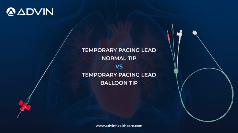
Temporary Pacing Lead – Normal Tip
The Normal Tip pacing lead is a traditional design that requires manual positioning within the heart. It provides stable pacing once correctly placed and is commonly used in controlled clinical environments.
Temporary Pacing Lead – Balloon Tip
The Balloon Tip pacing lead features an inflatable balloon at the tip, allowing it to float through the bloodstream and reach the desired position more easily. It simplifies placement, especially in emergency situations.
Quick Comparison Table
| Feature | Normal Tip Lead | Balloon Tip Lead |
|---|---|---|
| Placement Method | Manual positioning | Flow-directed (balloon) |
| Ease of Use | Requires skill | Easier placement |
| Procedure Time | Longer | Faster |
| Control | High | Moderate |
| Clinical Use | Planned procedures | Emergency use |
Key Feature Comparison
Normal Tip Lead Features
- Requires manual navigation and positioning
- Provides strong and stable placement
- Offers precise control during insertion
- Suitable for experienced operators
- Ideal for controlled clinical settings
- Reliable performance once positioned
Balloon Tip Lead Features
- Balloon-assisted, flow-directed placement
- Simplifies insertion process
- Reduces procedure time
- Useful in emergency situations
- Minimizes need for complex manipulation
- Allows smoother advancement through vessels
Common Features
- Used for temporary cardiac pacing
- Help maintain stable heart rhythm
- Compatible with standard pacing systems
- Designed for safe and effective use
- Suitable for hospital and critical care settings
- Sterile and single-use for patient safety
Key Differences
- Placement: Manual vs Balloon-assisted
- Ease: Skill-dependent vs Easier
- Speed: Slower vs Faster
- Control: High vs Moderate
- Use Case: Planned vs Emergency
Which One Should You Choose?
Choose Normal Tip pacing lead when
- Precise control and stable placement are required
- Procedure is planned and time is available
- Experienced operators are available
Choose Balloon Tip pacing lead when
- Quick placement is needed in emergency situations
- Ease of insertion is a priority
- Rapid patient stabilization is required
Conclusion
Both Normal Tip and Balloon Tip temporary pacing leads play a vital role in cardiac care. The Normal Tip offers precision and control, while the Balloon Tip provides speed and ease of use.
Selecting the right type based on clinical needs ensures effective pacing, improved patient outcomes, and efficient procedural management.
Get Connected:
+91-75037 27248 | cardiology@advinhealthcare.com | www.advinhealthcare.com