Virtual Reality Applications in Healthcare

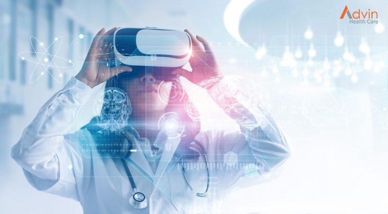
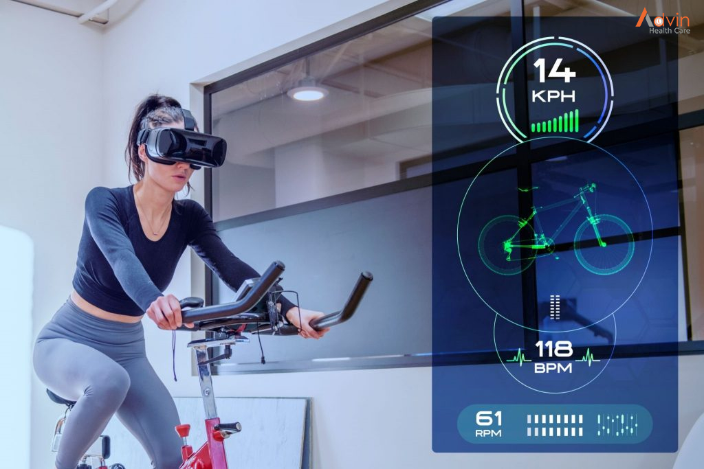
VR is the use of software to create an immersive simulated environment. Unlike traditional user interfaces, to experience VR, users put on head-mounted display (HMD) which places the user inside an experience, where they can engage with the environment and virtual characters in a way that feels real. VR has a unique power, more than any other technology that has ever existed, to make users believe they are in a different environment. This allows them to learn from experience as they would do in real life. This ability to deliver experiences on demand is where the power of VR lies.

Virtual reality in healthcare uses computer-generated technology for several healthcare applications, like providing virtual medical training to students and doctors or carrying out diagnoses, and many others.

There have been a lot of talks and discussions about the use of VR in the healthcare industry, with numerous applications of virtual reality already being implemented in healthcare. Applications such as robotic surgery, healthcare devices, etc. are slowly gaining popularity and are now more likely to be adopted by various healthcare organizations and accepted by the people. In this article we have talked about virtual reality and its applications in healthcare.

Applications of Virtual Reality in Healthcare


Medical Training

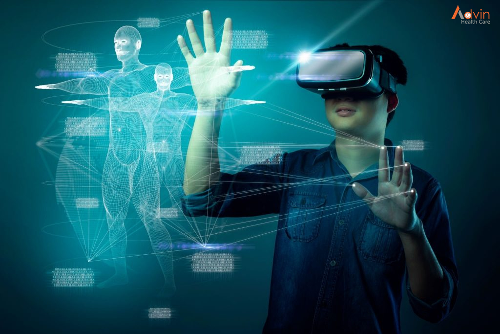
Virtual reality medical training is a modern method of training specialists in healthcare. This technology allows users to experience various scenarios in virtual surroundings. For example, users can immerse themselves by seeing a virtual human body from various angles and scales. Thanks to this, students and interns can train interactively at a higher level, while doctors can polish their professional skills.

VR is a technology that generates a virtual environment with an immersive effect. It is presented via a headset that includes VR glasses, headphones, and usually hand controllers for movement imitation, creating a sensation of reality. Therefore, users can experience situations they would never be able to in the real world because of physical limitations.

Virtual Reality in Diagnostics

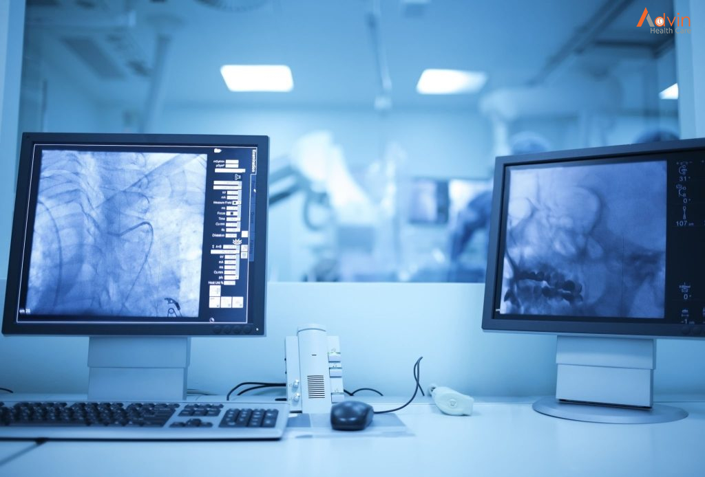
VR is being used as a powerful diagnostic tool, which helps doctors and physicians to carry out accurate diagnosis. This is done in combination with other methods, such as MRI/CT scans, and eliminates the need for any kind of invasive techniques, making it a pain-free experience for the patient.

Mental Illness Treatment

Exposure therapy is one of the standard procedures for treating different types of mental illness. VR is slowly changing the way exposure therapy is being carried out for mentally ill patients, by providing a low cost, flexible and low-risk solution to treat mental illnesses. By providing new ways to keep the body relaxed and calm, VR also helps in treating anxiety and panic attacks.

Pain Management

Cognitive distraction methods have been used by physicians to treat different types of pain for a long time now. VR provides a new face to these distraction methods by providing various interactive games. These games are played in a simulated environment and are loaded with interactive features. While some apps provide therapeutic VR for burn victims, there are also significantly advanced measures that facilitate efficient limb pain management. Furthermore, VR serves as a drug-free pain management alternative, which can be used with or without clinical care settings.

Physical Fitness and Therapy

The fitness industry is experiencing a huge change in the way exercises are carried out today. Many startups in the industry are combining cardio routines with VR and changing the way we exercise. Stepping further, VR also has a major role in physical therapy, where patients are subjected to an exercise routine that incorporates VR instead of drugs or invasive surgeries.

Human Simulation Techniques

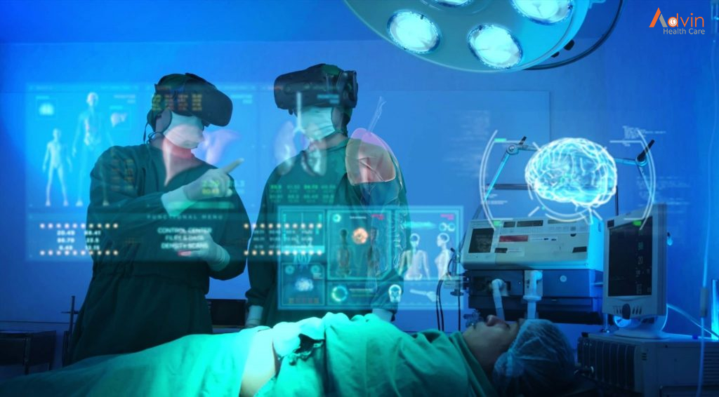
VR is very helpful in various human simulation techniques and enables doctors, physicians, nurses, and other medical professionals to interact with others. They can engage in different training sessions and interact with a patient, but only in a 3D environment. This is an immersive experience which determines the emotions of the participant using a series of sensors.

Top 5 Ways Technology Has Changed Dental Practices

Every day, oral healthcare providers must synthesize patient data such as intra- and extraoral exams, radiographs, clinical images and oral histories to develop treatment options. From there, a shared decision-making process between provider and patient serves to define the treatment plan, but the quality of the data and our ability to communicate and educate helps to inform patients’ decisions and shape the treatment path. The process of collecting patient data, documenting the clinical situation and planning treatment has been impacted by several key technologies.

The use of technology is no different. Tech advancement is usually created out of a need or a pain that is being addressed. As such, it is positioned to address those.

However, as you know, in life you cannot get anything without sacrificing something else. In the case of technology, you will be sacrificing both time and money. Time to learn the technology and its application and money to purchase the rights to use it ahead of your counterparts. This short article aims to put this sensitive balance in perspective and help you determine which technologies fit who you want to be as a dentist.

Considering the fallout of the COVID pandemic that is running rampant throughout the world, you have probably been exposed to hundreds of tech solutions that are being pushed with an increased sense of urgency. Having said that, the key is to remember that technology must enable your vision and purpose for your patients and not the other way around. The most important aspect of technology adoption is the understanding of the value received versus the price paid for the service.

We, understand that your ability to use the latest technology requires you to not only accept the cost of it, but to also ensure that you and your team are properly trained in using the new technology. It is imperative that you understand the benefits of pivotal technology and build it into your clinic plan. As we dive into the 3 main technologies to take advantage of in 2021.

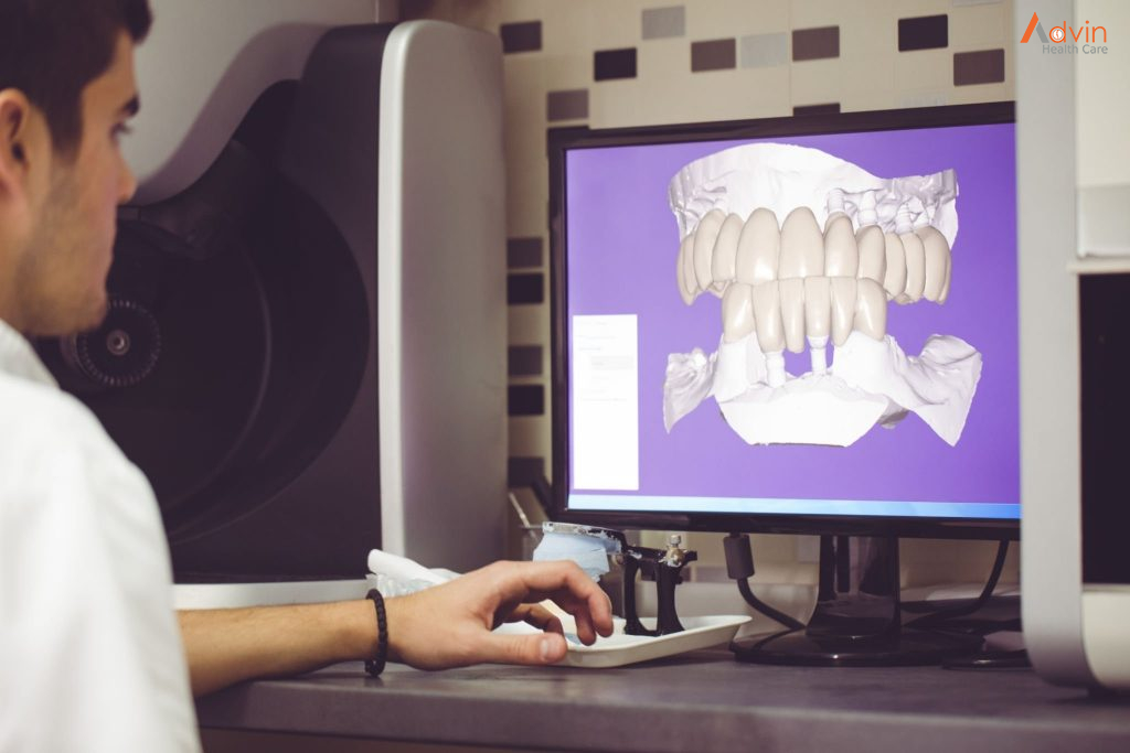
Digital Impressions


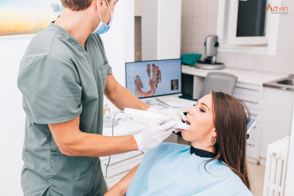
Traditional impressions have been one of the most obnoxious activities a patient can have in a dental clinic. A patient cannot have a positive experience in a clinic, if the impression material is sliding down their throat. Now that you had this unpleasant feeling We urge you to join the “digital dentistry” movement and ensure that you are working in a clinic that offers digital impressions and is incorporating digital tools to remove awkward experiences for patients.

We have rev  switched clinics. Not because they were performing bad dentistry, but because they were not using the most advanced tools to ensure my comfort. If no one is going to educate patients in your vicinity, you will be safe. However, I can guarantee that there are passionate and driven dentists in your vicinity who are planning and implementing strategies to win over these patients through education.

As the cheapest technology piece in this article, I can confidently say that the long-term benefits of having it outweigh the cost of both the system and the training required. In time, digital impression will become a standard of practice. Until then, you have the option of leveraging it towards building a strong patient base with patient comfort at the forefront.

Laser – Minimally Invasive Dentistry


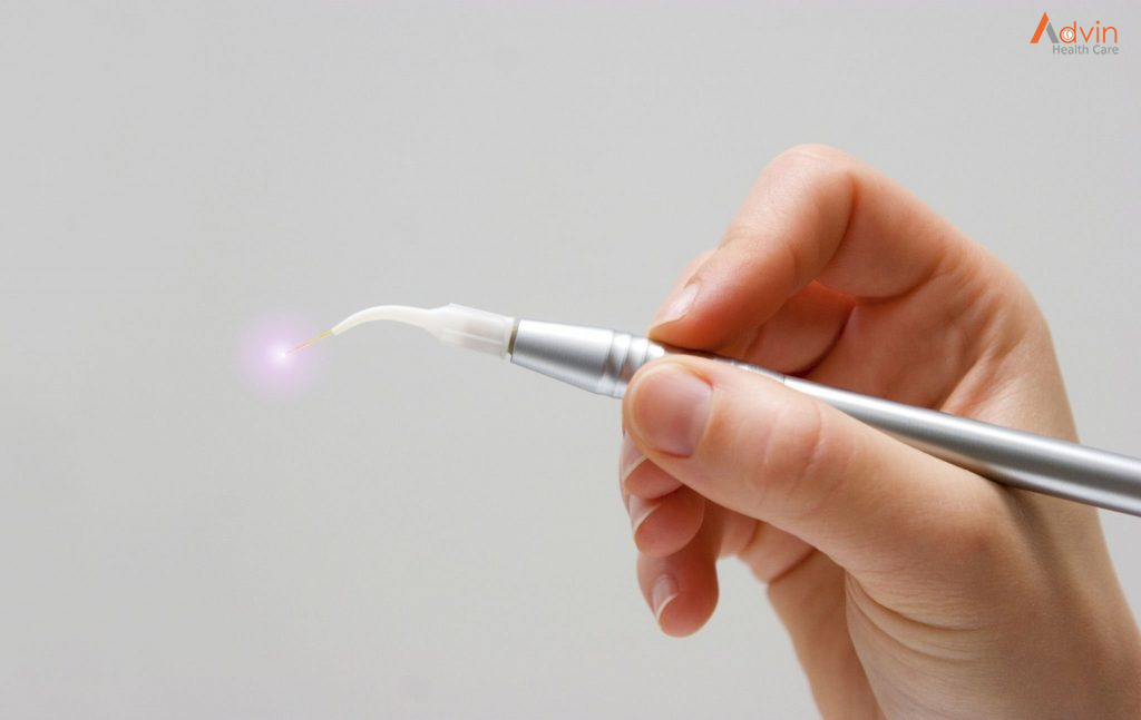
Minimally invasive dentistry (using a laser) has been around for quite a while now. The composers of this technology have failed to penetrate the market to the point of laser use becoming mainstream. Without getting into the politics of why that is, we like to urge you to explore this technology. The ability to treat conditions in a minimally invasive way with tools that are better than your metal instrument is another option you can offer your patients for conditions you traditionally just “watch”.

Considering that there are two main types of laser (soft and hard tissue), it is imperative you leverage industry training to ensure you profoundly understand this technology ahead of making it a unique provisioned service in your clinic. The versatility of laser technology in the treatment of a myriad of conditions is enough of an incentive to make it attractive to any young dentist in today’s industry.

As mentioned above, there are both soft and hard tissue lasers, which are grouped by their frequency. As you consider this technology, it is imperative you select the right vendor and the appropriate type of laser and education. This path requires a substantial investment as laser technology rapidly changes, but you can compensate for it by providing a wider array of services to your patients.

Digital Radiography


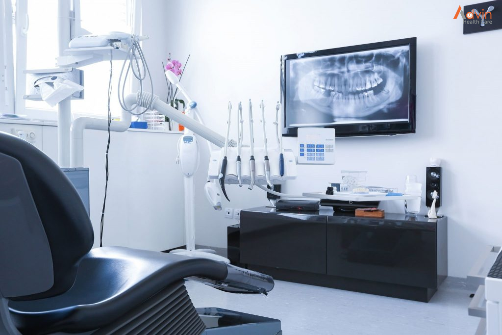
Digital capture of intraoral radiographs is simple, predictable and, just like loupes, has changed world. From the elimination of chemicals to the instant gratification of digital sensors and the ability optimize views on a large viewing monitor, digital radiography allows you to better diagnose potential problems. It is almost hard to believe that we were capable of appreciating early carious lesions on the small film we used to use!

Digital Scanning


The most recent technology is digital scanning. When combined with the electronic record, digital images and digital radiography, we can now not only educate patients about what their current situation is, but also show how we can address their complaints and even plan/predict the final outcome. For orthodontic, crown and bridge, and implant cases, we utilize digital scanning not only to plan treatment but also for communication between providers and laboratory technicians.

Digital Records


Perhaps it is because you run a software company that specializes in dental electronic records. Unfortunately, there are (too) many clinics that are still using paper charts for record keeping. After using a paper chart for over 20 years, electronic charts will be quick and efficient to document the patient’s needs. Having said that, the management and administration of recommended treatment through paper charts is a complete disaster.

If you are still reading, you agree that maintaining digital records is the way to go. Although this is true, and although colleges create a strict and clear outline of how records should be kept in a digital form, looking at the reality of record keeping, it appears that much is left to interpretation.

Digital X-Rays


There is much research out there with regards to digital radiations. Even if you prefer to take additional images, it will expose the patient to less radiation versus traditional x-ray equipment. The ability to have more angles of x-ray, while not exposing the patient to additional radiation, increases the probability rate of the acceptance and success of the proposed treatment. On top of digital x-rays, it is important to mention intra-oral images and fluorescent light images. Using these tools, you are better equipped to help patients with prevention of dental conditions.

Digital Charts


Let’s face it, you are looking for a quick way to document what you must so that you are able to continue caring for the patient. The problem is that the paper never translates into a database that easily. There is a substantial amount of manual entry by the existing administration team. Once you have transformed the information into digital patient files, you are able to continue and leverage the digital platform to create the appropriate treatment plan for the individual in question.

Oral Cancer

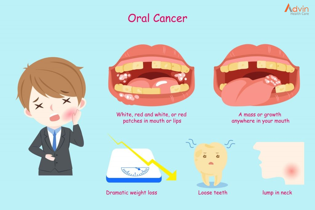
Mouth cancer refers to cancer that develops in any of the parts that make up the mouth (oral cavity). Mouth cancer can occur on the:

  • Lips
  • Gums
  • Tongue
  • Inner lining of the cheeks
  • Roof of the mouth
  • Floor of the mouth (under the tongue)

Cancer that occurs on the inside of the mouth is sometimes called oral cancer or oral cavity cancer.

Mouth cancer is one of several types of cancers grouped in a category called head and neck cancers. Mouth cancer and other head and neck cancers are often treated similarly.

Causes


Tobacco and alcohol use. Tobacco use of any kind, including cigarette smoking, puts you at risk for developing oral cancers. Heavy alcohol use also increases the risk. Using both tobacco and alcohol increases the risk even further.

HPV. Infection with the sexually transmitted human papillomavirus (specifically the HPV 16 type) has been linked to oral cancers.

Age. Risk increases with age. Oral cancers most often occur in people over the age of 40.

Sun Exposure. Cancer of the lip can be caused by sun exposure.

Symptoms


If you have any of these symptoms for more than two weeks, see a dentist or a doctor.

  • A sore, irritation, lump or thick patch in your mouth, lip, or throat.
  • A white or red patch in your mouth.
  • A sore throat or a feeling that something is caught in your throat.
  • Difficulty chewing, swallowing, or speaking.
  • Difficulty moving your jaw or tongue.
  • Swelling of your jaw that causes dentures to fit poorly or become uncomfortable.
  • Numbness in your tongue or other areas of your mouth.
  • Ear pain.

Diagnosis


Tests and procedures used to diagnose mouth cancer include:

Physical exam. Your doctor or dentist will examine your lips and mouth to look for abnormalities — areas of irritation, such as sores and white patches (leukoplakia).

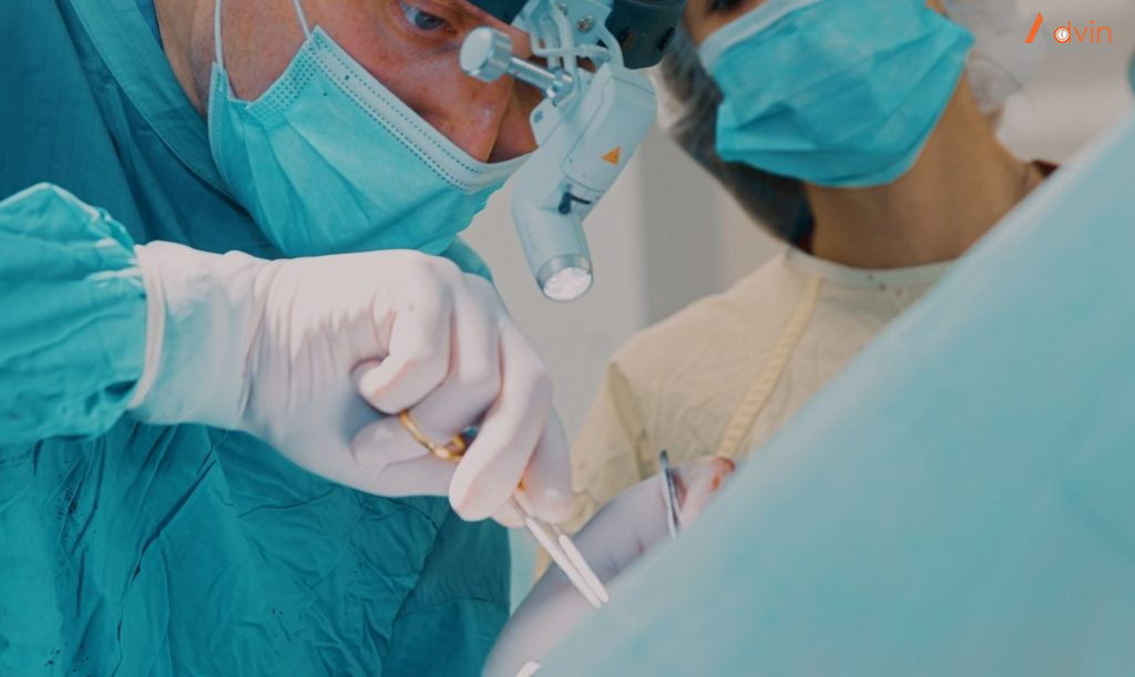
Removal of tissue for testing (biopsy). If a suspicious area is found, your doctor or dentist may remove a sample of cells for laboratory testing in a procedure called a biopsy. The doctor might use a cutting tool to cut away a sample of tissue or use a needle to remove a sample. In the laboratory, the cells are analyzed for cancer or precancerous changes that indicate a risk of future cancer. Determining the extent of the cancer

Once mouth cancer is diagnosed, your doctor works to determine the extent (stage) of your cancer. Mouth cancer staging tests may include:

Using a small camera to inspect your throat. During a procedure called endoscopy, your doctor may pass a small, flexible camera equipped with a light down your throat to look for signs that cancer has spread beyond your mouth.

Imaging tests. A variety of imaging tests may help determine whether cancer has spread beyond your mouth. Imaging tests may include X-ray, CT, MRI and positron emission tomography (PET) scans, among others. Not everyone needs each test. Your doctor will determine which tests are appropriate based on your condition.

Mouth cancer stages are indicated using Roman numerals I through IV. A lower stage, such as stage I, indicates a smaller cancer confined to one area. A higher stage, such as stage IV, indicates a larger cancer, or that cancer has spread to other areas of the head or neck or to other areas of the body. Your cancer’s stage helps your doctor determine your treatment options.

Treatment


Treatment for mouth cancer depends on your cancer’s location and stage, as well as your overall health and personal preferences. You may have just one type of treatment, or you may undergo a combination of cancer treatments. Treatment options include surgery, radiation and chemotherapy. Discuss your options with your doctor.

Surgery

Surgery for mouth cancer may include:

  • Surgery to remove the tumor.Your surgeon may cut away the tumor and a margin of healthy tissue that surrounds it to ensure all of the cancer cells have been removed. Smaller cancers may be removed through minor surgery. Larger tumors may require more-extensive procedures. For instance, removing a larger tumor may involve removing a section of your jawbone or a portion of your tongue.
  • Surgery to remove cancer that has spread to the neck.If cancer cells have spread to the lymph nodes in your neck or if there’s a high risk that this has happened based on the size or depth of your cancer, your surgeon may recommend a procedure to remove lymph nodes and related tissue in your neck (neck dissection). Neck dissection removes any cancer cells that may have spread to your lymph nodes. It’s also useful for determining whether you will need additional treatment after surgery.
  • Surgery to reconstruct the mouth.After an operation to remove your cancer, your surgeon may recommend reconstructive surgery to rebuild your mouth to help you regain the ability to talk and eat. Your surgeon may transplant grafts of skin, muscle or bone from other parts of your body to reconstruct your mouth. Dental implants also may be used to replace your natural teeth.

Surgery carries a risk of bleeding and infection. Surgery for mouth cancer often affects your appearance, as well as your ability to speak, eat and swallow.

You may need a tube to help you eat, drink and take medicine. For short-term use, the tube may be inserted through your nose and into your stomach. Longer term, a tube may be inserted through your skin and into your stomach.

Your doctor may refer you to specialists who can help you cope with these changes.

Radiation therapy

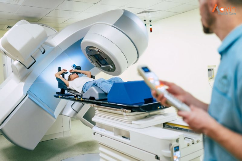
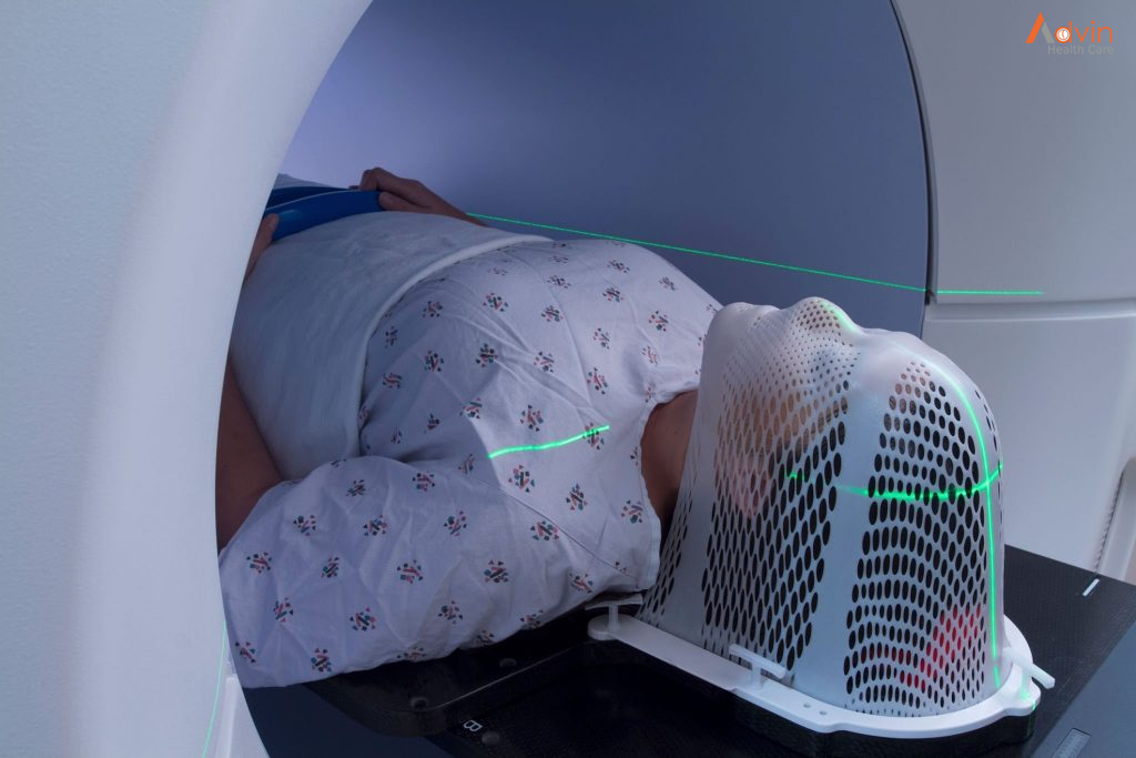
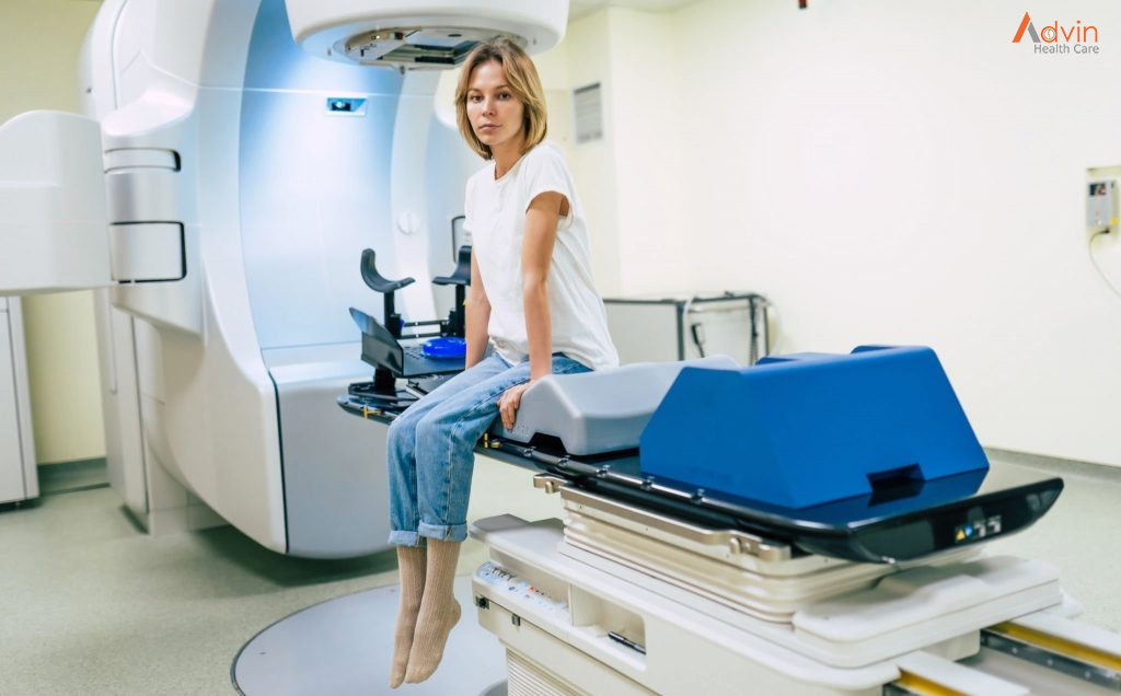
Radiation therapy uses high-energy beams, such as X-rays and protons, to kill cancer cells. Radiation therapy is most often delivered from a machine outside of your body (external beam radiation), though it can also come from radioactive seeds and wires placed near your cancer (brachytherapy).

Radiation therapy is often used after surgery. But sometimes it might be used alone if you have an early-stage mouth cancer. In other situations, radiation therapy may be combined with chemotherapy. This combination increases the effectiveness of radiation therapy, but it also increases the side effects you may experience. In cases of advanced mouth cancer, radiation therapy may help relieve signs and symptoms caused by the cancer, such as pain.

The side effects of radiation therapy to your mouth may include dry mouth, tooth decay and damage to your jawbone.

Your doctor will recommend that you visit a dentist before beginning radiation therapy to be sure your teeth are as healthy as possible. Any unhealthy teeth may need treatment or removal. A dentist can also help you understand how best to care for your teeth during and after radiation therapy to reduce your risk of complications.

Chemotherapy

Chemotherapy is a treatment that uses chemicals to kill cancer cells. Chemotherapy drugs can be given alone, in combination with other chemotherapy drugs or in combination with other cancer treatments. Chemotherapy may increase the effectiveness of radiation therapy, so the two are often combined.

The side effects of chemotherapy depend on which drugs you receive. Common side effects include nausea, vomiting and hair loss. Ask your doctor which side effects are likely for the chemotherapy drugs you’ll receive.

Targeted drug therapy

Targeted drugs treat mouth cancer by altering specific aspects of cancer cells that fuel their growth. Targeted drugs can be used alone or in combination with chemotherapy or radiation therapy.

Cetuximab (Erbitux) is one targeted therapy used to treat mouth cancer in certain situations. Cetuximab stops the action of a protein that’s found in many types of healthy cells, but is more prevalent in certain types of cancer cells. Side effects include skin rash, itching, headache, diarrhea and infections.

Other targeted drugs might be an option if standard treatments aren’t working.

Immunotherapy

Immunotherapy uses your immune system to fight cancer. Your body’s disease-fighting immune system may not attack your cancer because the cancer cells produce proteins that blind the immune system cells. Immunotherapy works by interfering with that process.

Immunotherapy treatments are generally reserved for people with advanced mouth cancer that’s not responding to standard treatments.

Lumbar Stenosis Pain

The term “stenosis” is derived from Greek and refers to the process of narrowing that constricts or “chokes” the spinal nerves. The cause of spinal stenosis in the lumbar spine (lower back) is commonly associated with degenerative changes, also known as spondylosis, that occur as a result of aging.

Lumbar spinal stenosis is a common cause of low back and leg pain, or sciatica.

As we age, the normal wear-and-tear effects of aging can lead to narrowing of the spinal canal, which houses the spinal nerves and spinal cord. This condition is called spinal stenosis.

Degenerative changes of the spine are seen in up to 95% of people by the age of 50. Spinal stenosis most often occurs in adults over 60. Pressure on the nerve roots is equally common in men and women.

A small number of people are born with back problems that develop into lumbar spinal stenosis. This is known as congenital spinal stenosis. Typically, this occurs in people who are born with a smaller spinal canal; because there is less space within the canal, degeneration, or arthritis, can affect them sooner. Congenital spinal stenosis occurs most often in men. People usually first notice symptoms between the ages of 30 and 50.

Spinal stenosis occurs when the space around the spinal cord and spinal nerves narrows. This puts pressure on the spinal cord and the spinal nerve roots, and may cause pain, numbness, or weakness in the legs.

Arthritis is the most common cause of spinal stenosis. Arthritis refers to degeneration of any joint in the body.

In the spine, arthritis can result as the disk degenerates and loses water content. In children and young adults, disks have high water content. As we get older, our disks begin to dry out and weaken. This problem causes settling, or collapse, of the disk spaces and loss of disk space height.

As the spine settles, two things occur. First, stress is transferred to the facet joints. Second, the tunnels through which the nerves exit (the foramen) become smaller.

As the facet joints experience increased pressure, they also begin to degenerate and develop arthritis, similar to that occurring in the hip or knee joint. As the facet joint wears down, the body responds by forming bone spurs to stabilize the joint. In addition, the ligaments around the joints that typically connect the bones together, called the ligamenta flava, increase in size. The combination of bone spurs and thickened ligaments crowds the space for the nerves, resulting in stenosis.

Symptoms


  • Back pain. People with spinal stenosis may or may not have back pain, depending on the degree of arthritis that has developed.
  • Burning pain in buttocks or legs (sciatica). Pressure on spinal nerves can result in pain in the areas that the nerves supply. The pain may be described as an ache or a burning feeling. It typically starts in the area of the buttocks and radiates down the leg. As it progresses, it can result in pain in the foot.
  • Numbness or tingling in buttocks or legs. As pressure on the nerve increases, numbness and tingling often accompany the burning pain, although not all patients will have both burning pain, and numbness and tingling.
  • Weakness in the legs or foot drop (difficulty lifting the front part of the foot).  Once the pressure reaches a critical level, weakness can occur in one or both legs. Some patients will have a foot drop, or the feeling that their foot slaps on the ground while walking.
  • Acute cauda equina syndrome. This rare condition is considered a medical emergency that requires prompt treatment. If the compression of the nerves is severe, you can experience numbness in you private area and lose control of your bowel and/or bladder. You may also lose strength in your legs and not be able to walk. If these symptoms occur, you may need emergency surgery.

Who does spinal stenosis affect?


Lumbar spinal stenosis primarily affects those over the age of 50. It has been shown that while nearly 50% of individuals over the age of 60 have lumbar spinal stenosis, not all cases require attention (Fritz et al., 2014). According to current research, there are three principal avenues that one can take to mitigate the degenerative condition that is known as spinal stenosis: physical therapy, epidural cortisone injections and decompressive surgery.

So I have spinal stenosis – what are my next steps?


For patients with spinal stenosis ranging from mild to moderate, physical therapy is the common route chosen. Even with patients suffering from severe spinal stenosis, it is usually recommended that they try some physical therapy before considering surgery. long with physical therapy, treating spinal stenosis through the use of epidural cortisone injections has become quite common. Containing a glucocorticoid and an anaesthetic, the purpose of the injection is to relieve pain by reducing the inflammation caused by the closing of the spinal canal or lateral recesses. The final path that one can choose for treating spinal stenosis is through decompressive surgery. It has been shown that the effectiveness of this method is higher the more severe the case.

How can Physiotherapy help?


Your physiotherapist can help determine which kind of stenosis you have (spinal or lateral).  They can provide you with education on your condition (including ways to help), some local manual therapy for your spine and corrective exercises to help improve your core strength and help you to move better.

Which exercises can I try to help?


We’ve listed a few different exercises to help.  Some are for general mobility, some are for strength.  If you have pain with any of these exercises, then stop and speak with one of our physiotherapists in Markham to help you with symptom relief and to get you moving better.

Knee to Chest stretch

Start in a lying position

Bring one knee to chest and hold for 30 seconds.  Repeat for the other side.

Knee to chest exercise

Cat-Camel; Cat-Cow exercise

Start in a four-point position.

Arch your back in one direction then move into the opposite arch.  Continue to move from one position to the other.  Try 8-12 repetitions.  There is no need to hold at the end.  There should be NO pain or tingling going down your legs – if there is, discontinue this exercise.

Hip Abduction 

Lie on your side. Bend your bottom leg.  Keep the top leg straight.

Lift your leg up towards the ceiling.  Hold for 1-2 seconds at the top, control the lowering.

These exercises are for guidance only.  Discontinue if there is any discomfort with these exercises and contact your physiotherapist for a detailed assessment and alternative exercises.

Types of Hospitals

A hospital is a healthcare facility that provides specialized medical and nursing care as well as medical supplies to patients. The most well-known form of the hospital is the general hospital, which usually carries an emergency department to handle urgent health issues such as fire and accident victims, as well as medical emergencies.

According to the hospital definition, a district hospital is usually the region’s primary healthcare facility, with a large number of intensive-care beds and extra beds for patients who need long-term care. Trauma centers, children’s hospitals, rehabilitation hospitals, hospitals, and seniors’ (geriatric) hospitals for coping with particular medical conditions such as psychiatric care are also examples of specialized hospitals and several other disease categories. When opposed to general hospitals, specialized hospitals can help save money on health care. Based on the source of revenue, hospitals are categorized as general, specialized, or government.

Types by Hospital size


One of the most obvious categories for comparing types of hospitals is the size of the institution. Hospital size is categorized based on how many beds it contains. The definition of what makes a hospital small, medium or large can vary depending on who you ask, but generally the lines break down into groupings similar to this:

  • Small hospitals contain fewer than 100 beds
  • Medium hospitals contain between 100 to 499 beds
  • Large hospitals contain at least 500 or more beds

Hospital size often corresponds to location. Rural hospitals tend to be much smaller, while larger hospitals generally only exist in urban areas. This shouldn’t come as a surprise—with a higher population density in urban areas, larger facilities are needed to accommodate the needs of the community.

Acute vs. Long-term care hospitals


Another potential distinction is whether a hospital specializes in long-term care. To be clear, the vast majority of all hospitals provide acute care. This means that they primarily care for patients dealing with illnesses, diseases, injuries, surgeries, surgery recoveries, obstetric care, postnatal care and trauma.

Long-term hospitals (LTCH) are often certified as acute-care hospitals, but also specialize in patients who on average, stay more than 25 days and are usually transferred from the ICU or a critical care unit, according to the Centers for Medicare and Medicaid Services. These patients often have more than one serious condition, and sometimes need to be treated with respiratory therapy, pain management treatments or for trauma to the head. Long-term patients often improve with time and care and are able to return home.

For-Profit vs. Not-For-Profit vs. Government-owned hospitals


Another potential differentiator between hospitals is the ownership structure. There are three primary options—For-profit, not-for-profit and publicly owned hospitals. Just over 51 percent of all registered hospitals are not-for-profit, according to the American Hospital Association. This means that they are not obligated to shareholders to earn profits—but that doesn’t mean they’re running a charity, either, as they need to remain financially viable. The biggest distinction is that excess funds typically remain in the hospital and can be invested in new technology, staff or research.

For-profit hospitals, on the other hand, are similar to other private businesses in that they are beholden to investors. Stakeholders demand a return on their investment, which can influence hospital operations. This means that there’s an increased pressure for for-profit hospitals to focus on efficiency in order lower costs. Some may concentrate on particularly profitable procedures and services in order to maximize profitability.

Publicly owned government hospitals are taxpayer-funded. In the U.S., these facilities are often associated with medical schools and do not have investors and quarterly profit projections to meet. That doesn’t mean budgets go out the window—there can still be challenges if public funding levels don’t keep up with patient demand and the needs of a facility.

Rural vs. urban hospitals


As mentioned in the size section, rural hospitals tend to be smaller and often fewer than 100 beds. Even more limited are “critical access” hospitals, containing fewer than 25 beds and are located 35 or more miles from the nearest hospital. These small hospitals depend heavily on Medicaid and Medicare funding to remain viable.

Urban hospitals are generally larger and have more competition as they are in densely populated areas with multiple hospitals for patients to choose from. Because of this competition, many urban hospitals specialize, either offering more versatility in treatment options and patient-experience perks.

Teaching vs. non-teaching hospitals


Teaching hospitals are affiliated with a university, medical schools or nursing schools. Interns, students and fellows work and learn at teaching hospitals while being supervised by qualified physicians, department leaders and other medical staff.

Non-teaching hospitals serve only the local community and don’t partner with any universities or medical schools. Does this make them better than teaching hospitals? Not necessarily. Teaching hospitals tend to treat sicker patients in order to provide more experiences to the students. Often these hospitals conduct academic medical research and have patients that take part in clinical trials designed to determine the effectiveness of new treatments. This can have a skewing effect on patient outcomes—many clinical trial participants seek out these less-proven treatments because their other options may be limited.

A teaching hospital may be a rewarding setting for experienced hospital staffers. Matthew Thomson RN, BSN, CCRN of The RN Mentor, has worked at two academic medical centres and says he loves the culture.

“What I enjoy the most about academic medical centres is the focus on learning,” Thomson says. “Attending physicians know that they are teaching the next generation of doctors. There’s a culture of mutual learning and respect.”

Federal hospitals vs. State and local hospitals


This classification of government-run public hospitals is based on where the funding comes from.  Federal hospitals are run and funded by the federal government include mostly Veterans’ Administration hospitals and clinics. The Department of Defense and the Department of Health and Human Services also run federally funded hospitals, but these hospitals aren’t commonly found in most communities.

Some hospitals are operated and funded by local state or county governments. These hospitals rely primarily on funding from local governments—though federal money via grants, Medicare and Medicaid payments certainly is part of their budget. These facilities are highly dependent on local funding—so they’ve seen some strain in states facing tough financial times.

Types of hospitals: Independent vs. hospitals in a system

Many hospitals are a part of a system that can include one or multiple hospitals and several clinics and separate specialized facilities. This allows for more comprehensive outpatient care once a patient leaves the hospital or emergency room. Independent hospitals that are not a part of a system are becoming rare as mergers are becoming more common.

Independent hospitals and hospitals in a system can both be a part of a healthcare network— a group of healthcare facilities, physicians, insurers, agencies and others that work together to coordinate services. Though they have separate administration systems, they collaborate for the community’s benefit.

Trauma levels hospitals


In addition to being classified by funding and function, hospitals are also evaluated based on their readiness to treat trauma resulting from accidents, falls, gunshot wounds or other massive injuries. Though criteria for the trauma categories vary from state to state, there are common denominators. Let’s take a look at the polar ends of the trauma center scale to get a better idea of what these designations mean.

Level I trauma centers can provide total care, from prevention through rehab, for traumatic injuries. They provide 24-hour in-house coverage by general surgeons, and specialists like orthopedic surgeons, neurosurgeons, plastic surgeons, anesthesiologists and other critical-care providers. The hospital has to meet a minimum requirement of seriously injured patients in their care each year. In addition to providing top notch and prompt care, they also provide public education and leadership in prevention while being a referral resource for community members in the area, according to the American Trauma Society.

On the other end of the trauma center spectrum, Level-V centers can provide initial evaluation, stabilization and diagnostic capabilities by one or more trauma nurses and physicians before transferring patients, if needed, to Levels I through III trauma centers that they have agreements with. Level-V centers may be able to provide surgery and critical-care services, but it is not guaranteed.

Specialty hospitals


Outside of these primary categorizations, there are many other types of hospitals, usually specializing in certain types of care. Here are three common examples of specialty hospitals:

  • Children’s hospitals focus on conditions and diseases that affect young patients. The staff at these hospitals are specially trained to work with children and are able to offer psychosocial support for the children who require extended stays.
  • Research hospitals are committed to discovering cures for certain conditions while treating various illness and diseases.
  • Psychiatric hospitals specialize in mental health needs and illness using medication, psychotherapy and behavioural therapies. Some focus on short-term, emergent treatment, while some guide patients through long-term stabilization.


Important Of Disposable Products

According to the Food and Drug Administration (FDA), the governing entity in the United States that regulates medical devices, a single-use or disposable medical device is “intended for use on one patient during a single procedure. It is not intended to be reprocessed (cleaned, disinfected/sterilized) and used on another patient. The labelling may or may not identify the device as single use or disposable and does not include instructions for reprocessing.” The disposable medical supplies industry produces supplies you frequently see when you interact with a medical professional – bandages, hypodermic needles, exam gowns, gloves, catheters, surgical sponges, tongue depressors, syringes, pregnancy and drug tests, and face masks. Consumable medical devices are like disposable devices in that they also ultimately should be disposed of but can be used more than once or for a length of time. These include devices like elastic bandage wraps, pacemakers, and drill bits and blades for surgical instruments.

At Advin, we work with medical device use in Hospital of all shapes and sizes – small and large, start-up and established – that develop and manufacture a wide array of devices. No matter how varied the companies are, across the board their biggest concern is keeping patients safe. When designing a medical device, companies want to do everything they can to improve medical device UX, or user experience, and reduce the risk of harming their user. Disposable medical devices can help.

Anytime device companies can incorporate disposable elements or create entirely disposable medical products, they are improving patient safety and reducing risk. The primary reason is that using disposable medical supplies and devices prevents cross contamination between patients. Reusable medical devices must be completely and thoroughly sterilized before they can be used again. In addition to expanding design considerations for sterilization, companies are subject to increased regulatory responsibilities. The FDA has strict sterilization process controls; devices must prove they adhere to in the submission process. The role of sterilization and disposable medical devices in preventing the spread of disease has increased in relevance during the COVID-19 pandemic.

Not being able to properly sterilize a medical device can have serious consequences. In 2013, the Centers for Disease Control and Prevention (CDC) alerted the FDA of a potential connection between duodenoscopes, devices that pass into the small intestine, and antibiotic-resistant infections, even though users reported following proper cleaning and disinfection or sterilization protocols outlined by the manufacturer. Incidences such as these have led companies to design disposable elements like covers for non-disposable medical instruments.

Another critical benefit of disposable medical devices is durability, especially when it comes to consumable devices. By their nature, some devices, or individual components of the device, wear out after a certain period – lights burn out, batteries die, drill bits and blades dull, sensors break, and wires fray. This is also where the importance of testing and labelling comes in. Medical device companies must test to identify the average number of uses a device or component can withstand while working effectively and label the device accordingly to indicate how many times it can be used within the indicated safety margin. At

Disposable medical devices, or disposable components of devices, can help improve the health and safety of the patients we serve. That’s why, when we work with our clients on their medical device design, development, and regulatory submission, we look for opportunities where incorporating disposable elements makes the system safer. If any of our services can support your product, including our medical device prototype development.

Disposable or single-use medical devices are the ones that can only be used once and then needs to be discarded. When it comes to offering efficient healthcare services, hygiene and sanitation are the two important factors. Despite having a multitude of sterilization methods like chemical sterilization, heat sterilization, etc, there is always a scope of contamination left behind. And in this covid era, the transmission of infection is the first thing one needs to get rid of. Hence, the only way that absolutely eliminates the chance of contamination is the use of disposable products. Almost every industry is now switching to the use of single-use products as much as possible. Therefore, it becomes an essential choice for the healthcare industry.

Benefits of Disposable Medical Supplies


Here are some of the most common benefits that can be attained with the use of disposable medical products:

Prevention of infection – Research was conducted to investigate the presence of microbes on disposable and reusable medical products. More than 30 percent of the wet swabs taken from reusable products contained pathogens while most of the wet swabs taken from the disposable products were found sterile. In fact, the Center for Disease Control and Protection states that the use of disposable products improves patient safety by eliminating the risk of patient-to-patient contamination.

Saving time – The reusable products need to be cleaned and sterilized after every single use. On the other hand, single-use products are mostly ready to use and can be disposed of after use. This saves a lot of time for the medical practitioners and increases their efficiency.

Cost-cutting – Even though it may seem like an expensive choice to most practitioners, single-use products are in fact more cost-effective in the long term. This is because reusable devices have a high cost which further increases due to the need of purchasing additional accessories. Furthermore, the products require regular cleaning and maintenance. The extra cost is added for their regular sterilization.

On the other hand, single-use products are much cheaper, need the least maintenance, and do not require extensive sterilization procedures. There are no additional costs associated with the repair. Instead, they are capable of providing similar quality as that reusable products.

Impact on the environment – It may seem to many that disposable products have more detrimental effects on the environment, but this is not the case. If disposed of properly, the single-use products are environmentally-friendly. On the other hand, the waste produced in the form of chemicals and air pollutants while manufacturing reusable medical devices is much more harmful to the atmosphere.

Modular Operation Theatre

In the operating department for which the most integrated function is required, cleanliness must be kept and the safety of facilities, equipment and devices must be maintained any times. Furthermore, the working environment for medical staff such as doctors and nurses must be considered from the viewpoint of human engineering. Our modular operating theater satisfying those conditions consists of wall, ceiling and slanted panels, and is capable of incorporating not only electrical equipment, medical gas system, and lighting gears but all the necessary functions and equipment at need. In addition, the modular theater has a high availability of extension providing for the introduction of a new facility in the future, establishing as well an operating environment with high workability and enough function.

Operation theatre is that specialized facility of the hospital where life-saving or life-improving procedures are carried out on the human body by invasive methods under strict aseptic conditions in a controlled environment by specially trained personnel to promote healing and cure with maximum safety, comfort, and economy. It is imperative that operation theatre (OT) is designed scientifically to ensure sterility, easy maintenance, and effective utilization. A civil – mechanical – electrical – electronic – biomedical combination driven and coordinated by needs, preference and safety of the medical-surgical team form the basis of planning, starting and maintaining an operation theatre.

What is a modular operation theatre?


It is a compact and highly sterilized setup containing particular air filters to control the airflow system within. Some specific types of air conditioning and air handling units are used to maximize protection against contaminants to increase the physical safety of patients. The walls and ceilings are made of pre-engineered panels and the floors are chemical resistant and antistatic making the operation theatre easier to install and clean.

Why do we need Modular OT?


Sterility: To have complete infection free cases

  • To achieve a very low level of Bacteria count in the ot
  • To have non-porous, seamless, easily cleanable surfaces
  • To reduce the need of frequent disinfestation.

Work-Flow Management

  • To have concealed wiring and medical pipeline system
  • To have a complete hassle-free floor area leading to enhanced equipment and personal movement
  • To have effective lighting system to operate under better conditions.

Aesthetics – Pleasing work environment

  • Effective lighting by maintaining LUX levels
  • Clean air quality and effective ventilation
  • Pleasing work environment through the use of different walls and floor colours.

Features of a Modular OT?


  • A continuous flow of highly filtered “Bacteria free” air into the operation theatre.
  • A continuous removal of contaminated air out of the operation theatre
  • Durable anti-bacterial paint
  • Easy to clean and maintain
  • Permeability of vapour to escape which protects against the growth of yeast and microbes
  • Moisture and Thaw resistance
  • Easy to move and get hands on the instruments
  • Should be able to accommodate a number of doctors inside.

Major components of Modular OT system


The Wall and ceiling

A modular OT is a free-standing structure constructed inside a hospital room. The panels of Modular OT are made of Steel or glass. A 200 micron coat of anti-microbial paint is sprayed on the inside of the panels facing the OT table.

All four corners of the room will have provisions for air duct. Sufficient room should be given inside the operation theatre for easier operation procedure and the remaining space should be used for storage. The joints and cavities must be filled with epoxy and sanded flush to provide a joint less surface.

Laminar air flow unit – Plenum

A Modular OT consists of a laminar air flow unit which supplies air into the Modular OT. Laminar flow is defined as the flow of fluid in which the entire flow is steady with uniform velocity in which the flow of lines is parallel to each other.

Laminar flow unit is used in Modular OTs

  • To control the temperature and humidity of space,
  • To assist in the removal and dilution of waste aesthetic gases
  • To dilute airborne bacterial contamination
  • To control the movement of air within the department, such that the amount of airborne bacteria is minimal.

Light

Lights of a Modular OT are fixed to its ceilings. Clear and effective lighting is crucial to the operation. Integral lighting systems provide illuminations in excess of 1300Lux.

Pendants 

Pendants are used in Modulate OTs for convenient positioning of Medical gas pipeline, Equipment and other tools. These are flexible enough to be fixed at variable heights and distances.

Pendants provide,

  • Medical gas supply
  • Electrical power supply
  • Data points
  • Shelves for some equipment
  • Drawers for accessories
  • Mounting of medical devices and others

Hermetically Sliding door

The use of hermetically sliding door is to maintain the correct air pressure inside the modular OT. Also Hermetically sliding doors use very less space compared to hinges or any other types of doors. The door should have rubber wheels and made to run on a nylon platform to reduce the abrasion. The doors can be either automatic with controls or manual. It should also be worked manually at the time of power failure.

OT Control panel

OT Control panel is an electric control panel fixed to the wall of modular OT. Nearly 9 types of controls can be mounted on the panel. These controls include medical gases, medical equipment, Ventilation system etc…

Anti-Static Flooring 

Anti-static flooring is used in industries where static can cause interference or damage to employees and/or equipment. Anti-static industrial floors inhibit the generation of electrostatic discharge or ESD, which is the pulse of static electricity that happens when a charged person or surface comes into contact with another object.

Stroke Rehabilitation

With the thoughts that are shifting between hope and anxiety, the stroke survivors and their family members go through a lot. This happens especially after the discharge from the hospital.

While a few survivors recover on their own, at least two thirds of stroke survivors need some sort of rehabilitation. The extent of the therapy is determined by the physiatrist who is experienced in Stroke Rehabilitation.

The initial consultation followed by a thorough analysis of the current condition of the survivor, the therapist sets up the goal, and devises a therapy plan upon consulting other rehab specialists.

This therapy could be typically availed as an outpatient service but few facilities offer it as an inpatient rehabilitation service. The roadmap to therapy, and eventually to recovery, is laid after considering every minute aspect that can pose a threat or cause an interruption during the therapy.

Setting up a platform that works seamlessly


The goals are set, the path is fixed, and the rehabilitation is started. But what to expect from the whole therapy course?

The rehabilitation experts, as a team or as individuals, start working towards the time-bound goals. The progress will be evaluated on a weekly basis and the pace is checked against the date.

If there are any challenges related to the speed of recovery, responsiveness, involvement of the survivor, these weekly evaluations address them timely and try to get the recovery back on track.

Communication, and an empathetic and supportive environment play an important role during the recovery. And, it is possible only when the team of doctors, patients, and their family members align themselves to the determined goals.

What to expect from the stroke rehabilitation therapies?


Each stroke survivor will have different sets of needs and different therapists work on addressing them. Typically here is what to expect from the therapy sessions.

  • Therapy sessions – Employing both the individual and group therapy sessions whenever there is need, the rehabilitation specialists conduct therapy sessions on a daily basis. Again, the frequency of these sessions is determined by the physical medicine and rehabilitation experts and varies from person to person.
  • Support just the way it is needed – Positive approach in a therapy always yields better results and the survivors feel positive when they feel they are being recovered in a supportive and engaging environment. Expect psychological and emotional support from the therapists, communicate with them, and express the difficulties or discomforts.
  • Occupational therapy – The stroke survivors need additional strategies and comfortable ways to go about with the day-to-day activities and the experienced and trained occupational therapists will help them manage their day in a more comfortable way. This support may also be extended to the career and professions.
  • Using the techniques such as neuroplasticity – Stroke survivors might have to learn the skills even though they knew it in the pre-stroke days. The brain needs to make new connections, and learn skills again. It takes time, practice and most importantly patients and determination.
  • Therapy support from the other therapists – Stroke can lead to complex medical conditions such as vision and speech impairments. Vision therapy, and speech therapy for stroke patients is aimed to help the person to perform these functions in a better way.
  • Support in physical activities – Everyday physical activities are essentials in the process of therapy. The therapists should help the survivors in performing these activities and these sessions should be in a one-on-one environment.

Therapy in a most holistic, advanced, supportive, and collaborative environment. In a nutshell, this is what the survivor or the family member should expect from stroke rehabilitation.

The importance of building trust and nurturing relationships between the survivors and the therapy team could not be emphasized further. This could be the backbone during the entire process of rehabilitation therapy for stroke survivors.

Radiology Tests Explained

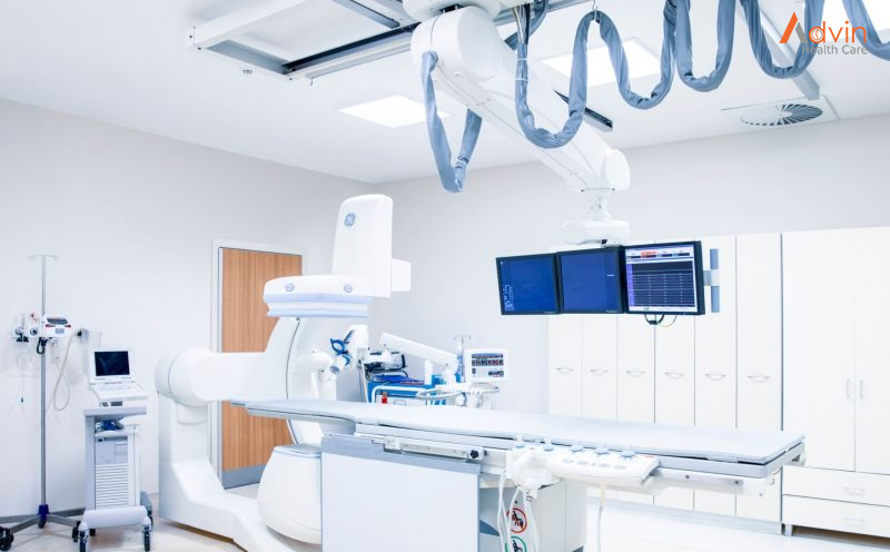
Radiology defines a wing of medicine that functions using radiant energy while diagnosing and treating diseases. In simple words, radiology means a process to determine whether a medical condition is present or not before moving ahead with the treatment. It is a test that takes images of different body parts and tries to find relevant issues to support a necessary diagnosis.

This branch of medicine comprises two areas – diagnostic radiology and interventional radiology. Both these forms use different radiant energy to provide a required diagnosis. Among the various radio imaging exams, some of the most common include- MRI, Ultrasound, X-ray, PET scan, etc. After the patients experience these tests, the radiologist will provide reports of their elucidations to the concerned doctors.

Now that individuals have a fair understanding of what radiology is, they should know about this diagnostic test’s different types and procedures.

The five most common radiological tests prescribed by doctors are explained below.

X-Rays


X-ray is the most common and widely performed radiological test. It is non-invasive and painless. This method makes use of electromagnetic radiation to detect even minor anomalies. Modern x-ray machines have become more efficient, with a ray source on one side and radiation-sensitive plates on the other. It helps in quick imaging and gives instant results.

The Test Procedure

The patient has to place the concerned body part between the ray source and the radiation plate. The doctor might ask them to move or turn to take images from different angles. This process takes only about 10 to 15 minutes.

Simple x-rays are done for screening as well as diagnostic purposes. It can:

  • detect hairline fractures, ligament tears, or lumps in certain areas
  • diagnose conditions like osteoporosis, arthritis, digestive tract issues, and infections
  • track the severity of an injury and the effects of treatment on the patient’s body
  • screening for and diagnosing the malignancy of breast lumps (mammogram)
  • live imaging of blood flow, hard and soft tissues, and joints using a contrast dye like barium (fluoroscopy/barium swallow radiology)

Ultrasound


Ultrasound, also called sonography, is another widespread radiological procedure. It uses high-frequency sound waves to detect different structures inside the body. There are no radiation waves in this test which makes this a safe procedure even for pregnant women. Ultrasound can be minimally invasive or non-invasive and mainly studies soft tissue structures and organs.

The Test Procedure

The doctor applies a gel on the target body part and places a transducer or probe which emits ultrasound waves. They move the probe around the body part to get a clear picture. For endoscopic ultrasound, the probe is inserted into the body through the patient’s mouth or rectum, depending on the part to be studied. The procedure can take about 25 minutes to 1 hour.

Purpose Of The Test

Ultrasound is used for diagnostic and interventional purposes. It is performed to:

  • diagnose breast lumps, joint inflammation, and issues related to gall bladder and prostate
  • diagnose inflammation or cancer in the digestive tract
  • study the progression of a pregnancy
  • act as a guide during biopsies

CT/CAT Scan


A computerised Axial Tomography scan uses a series of ionising radiations to form a cross-sectional image of the patient’s whole body. It is a non-invasive procedure and helps study almost all body structures.

The Test Procedure

The patient is made to lie on a table that slides into the doughnut-shaped scanner. A scanning tube at the centre rotates around the patient’s body and creates a 3-D image. There is a transmitter inside the scanner through which the patient can talk to the doctor in case of nervousness or discomfort. The imaging takes only 15 to 20 minutes.

Purpose Of The Test

Doctors usually prescribe a CAT scan when other tests indicate some abnormality. It is done for diagnostic and radiology treatment purposes like:

  • studying the severity of a traumatic injury or fracture
  • detecting tumours and other abnormalities
  • diagnosing infections and heart and vascular diseases
  • guiding biopsies

MRI Scan


Magnetic Resonance Imaging works very similarly to a CAT scan. Only, it uses magnetic fields instead of ionising radiation. This test works for almost all body parts and conditions.  Due to the absence of radiation, it is also considered a safer method. However, it takes longer than a CAT scan.

The Test Procedure

The patient is made to lie on the sliding table, which moves into the MRI scanner. The scanner rotates, sending magnetic waves to create a 3-D image of the patient’s body. As it does so, the scanner makes loud noises. The patient can interact with the doctor through the transmitter in case of discomfort.

Purpose Of The Test

MRI scan helps in diagnosing a wide array of conditions. It includes:

  • spinal cord diseases
  • issues related to soft tissues like organs, blood vessels, joints, and tendons
  • strokes, aneurysms, and multiple sclerosis

PET Scan


Positron Emission Tomography scan follows a different methodology than the ones discussed above. In this test, a radioactive drug, called a radiotracer, is inserted into the patient’s body. The PET scanner studies the transmission of this tracer on a cellular level. This radiography test helps study any abnormalities and the functioning of different body systems.

The Test Procedure

The radiotracers are introduced into the patient’s body via an injection into the veins or inhaling gases, or drinking a mixture containing radiotracers. It depends on the organ system or body part to be studied. After about an hour of introducing the radiotracer into the body, the scanning is done. The whole procedure might take up to  2 hours. The radiotracers are introduced into the patient’s body via an injection into the veins, inhaling gases, or drinking a mixture containing radiotracers.

Purpose Of The Test

PET scan is a highly efficient procedure for performing diagnostic imaging. It is used to diagnose:

  • cancer
  • cardiovascular diseases
  • Alzheimer’s disease
  • Parkinson’s disease
  • digestive tract diseases
  • seizures
  • epilepsy

Future Technology in Orthopedic

Virtual reality, sensors and 3D printing for a better follow-up of the patient throughout his journey


If virtual reality can already be used to treat certain phobias, or even to help with rehabilitation thanks to mirror neurons, for example, the application of this technology combined with sensors and 3D printing could improve patient care throughout the course of their treatment, from diagnosis to post-operation follow-up.

Before the orthopedic operation and as a preventive measure, sensors could be used to monitor the patient’s physical data. For example, we could imagine a custom-made corset, which integrates sensors to follow in real time the evolution of the curvature of the back, as well as the use of post-operative implants capable of detecting possible complications.

Virtual reality technologies can also be used to plan and simulate the operation to be performed in a virtual environment, allowing the patient to experiment with several procedures in order to select the most appropriate one.

Virtual and mixed reality for the learning and training of doctors in orthopedics


Originally, virtual reality in health was developed for educational purposes, for example by allowing a 3D reconstruction of the human body, to study in detail the different components and mechanisms of the body. This technology allows, for example, reconstruction by organ, and access to high quality educational information.

The development of virtual reality is revolutionizing surgical training methods: little by little, traditional methods such as teaching in the operating room or cadaver labs are being abandoned in favor of virtual reality, which provides a better viewpoint and a better understanding of the manipulations to be performed. This trend has become even more pronounced since the Covid-19 crisis, during which virtual reality has made it easier to train future surgeons.

Mixed reality, on the other hand, allows access to an immersive tutorial during an operation, to ensure that the correct actions are repeated.

Robotics and augmented mixed reality for greater precision in the orthopedic surgery operating room


By introducing robotics, as well as augmented and virtual reality in the operating room, it is possible to significantly reduce variability during operations, to guarantee better results for patients.

The use of mixed reality in the operating room for orthopedic surgery, allowing the surgeon to be faster and more precise, is more recent. It was notably Prof. Thomas Gregory, in 2017, who demonstrated the interest of this technology in the operating room for orthopedics. Indeed, he operated on a shoulder prosthesis, assisted by mixed reality glasses, projecting a digital model on the patient, and gathering all the information collected during imaging examinations. The information available included, for example, the exact thickness of the tissue, or the precise location of surrounding organs invisible to the naked eye.