Introduction: Visualization and Comfort in Cystoscopy
Cystoscopy is a fundamental procedure in urology used to examine the urethra and bladder for diagnostic and therapeutic purposes. The choice between a Rigid Cystoscope and a Flexible Cystoscope significantly impacts patient comfort, procedural efficiency, and clinical outcomes.
While both instruments serve the same purpose, they differ in design, maneuverability, patient tolerance, and application settings.
Product Overview in Clinical Use
Rigid Cystoscope
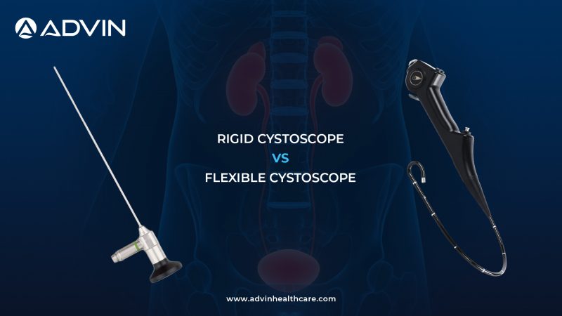
The Rigid Cystoscope is a straight, non-flexible instrument commonly used in standard cystoscopic procedures.
Key characteristics:
- Straight metal construction
- High-definition optics
- Larger working channel
Clinical advantages:
- Superior image clarity
- Better instrument control
- Ideal for therapeutic interventions
Flexible Cystoscope
The Flexible Cystoscope is designed with a bendable shaft, allowing it to navigate the natural curvature of the urinary tract.
Key characteristics:
- Flexible, steerable tip
- Smaller diameter
- Advanced fiber-optic or digital imaging
Clinical advantages:
- Increased patient comfort
- Easier navigation through urethra
- Suitable for outpatient procedures
Workflow-Based Usage: Control vs Comfort
Both cystoscopes are used for bladder visualization, but their usage differs based on clinical needs:
- Rigid Cystoscope → Better control for procedures and interventions
- Flexible Cystoscope → Enhanced comfort for diagnostic examinations
This affects:
- Patient experience
- Procedure type
- Clinical setting
Core Differences That Drive Clinical Choice
The key difference lies in flexibility and patient tolerance:
- Rigid cystoscopes offer precision and stability
- Flexible cystoscopes provide comfort and adaptability
Quick Comparison Overview
| Aspect | Rigid Cystoscope | Flexible Cystoscope |
| Structure | Straight | Flexible |
| Patient Comfort | Moderate to low | High |
| Image Quality | Excellent | Good to excellent |
| Maneuverability | Limited | High |
| Working Channel | Larger | Smaller |
| Procedure Type | Diagnostic + therapeutic | Mostly diagnostic |
| Setting | OT / hospital | OPD / outpatient |
| Cost | Lower | Higher |
Clinical Preference & Real-World Application
Rigid Cystoscope is commonly used in:
- Therapeutic procedures (biopsy, stone removal)
- Operating room settings
- Situations requiring larger instruments
Its stability and clarity make it ideal for interventional work.
Flexible Cystoscope is preferred in:
- Routine diagnostic cystoscopy
- Outpatient (OPD) settings
- Patients requiring minimal discomfort
Its flexibility improves patient experience and procedural ease.
Practical Selection Guide
- Choose Rigid Cystoscope for therapeutic and surgical procedures
- Choose Flexible Cystoscope for diagnostic and outpatient use
- Use rigid when control and instrumentation are required
- Use flexible when patient comfort is a priority
Conclusion: Balancing Precision and Patient Comfort
Both rigid and flexible cystoscopes are essential in urology:
- Rigid Cystoscope → Precision, clarity, and control
- Flexible Cystoscope → Comfort, flexibility, and ease of use
Modern urology practices often use both instruments together to ensure comprehensive patient care.
Why Advin Health Care is the Right Choice
Advin Health Care offers a complete range of cystoscopy solutions designed for precision and reliability.
- Rigid cystoscopes deliver excellent visualization and procedural control
- Flexible cystoscopes provide advanced navigation and patient comfort
With Advin, healthcare providers benefit from:
- High-quality instruments
- Durable design
- Consistent performance across all cystoscopic procedures
Get Connected:
+91-70717 27261 | urology@advinhealthcare.com | www.advinhealthcare.com