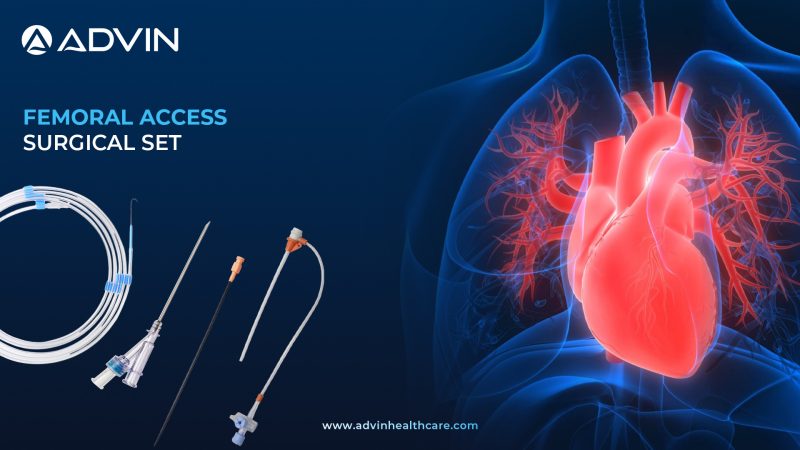
Pancreatic Necrosectomy – Surgical Management of Pancreatic Necrosis

Pancreatic Necrosectomy Intro

Pancreatic Necrosectomy is a surgical procedure used to remove dead or infected pancreatic tissue resulting from severe acute pancreatitis. It is usually performed when necrosis leads to infection or organ dysfunction. This procedure helps improve survival and reduce complications in critically ill patients.

Precise Surgical Removal of Necrotic Tissue

Pancreatic necrosectomy involves accessing the necrotic area through open surgery, minimally invasive techniques, or endoscopic approaches. Surgeons carefully remove the dead tissue while preserving as much healthy pancreas as possible. In many cases, endoscopic transgastric necrosectomy is performed using a flexible endoscope and specialized tools to debride the necrosis through the stomach wall. Alternatively, video-assisted retroperitoneal debridement (VARD) may be used in selected cases. Drainage catheters are often placed to remove residual fluid collections and prevent further infection. This step-by-step removal reduces inflammation and supports the patient’s recovery while minimizing damage to surrounding organs.

Key Medical Indications for Surgery 

  • To treat infected pancreatic necrosis unresponsive to antibiotics
  • To relieve pressure and inflammation caused by large necrotic tissue collections
  • To prevent life-threatening complications such as sepsis or multiorgan failure
  • To manage persistent symptoms like fever, abdominal pain, or vomiting

Products used in Pancreatic Necrosectomy

  • Endoscopic Ultrasound (EUS) Scope
  • Cystotome
  • Lumen-Apposing Metal Stent (LAMS)
  • Biliary Dilator
  • Necrosectomy Forceps
  • Endoscopic Drainage Catheter
  • Irrigation Suction Cannula
  • Guide Wire
  • Stone Retrieval Basket 
  • Nasocystic Drainage Tube

Countries with High Surgical Expertise 

  • United States
  • Germany
  • Japan
  • India
  • South Korea

Clinical Advantages of Tissue Debridement 

  • Effectively eliminates infected pancreatic tissue
  • Reduces hospital stay and ICU complications
  • Improves survival rates in severe pancreatitis
  • Allows for minimally invasive treatment in selected cases
  • Decreases the risk of recurrent fluid collections or organ damage

Advin Health Care is a globally recognized provider of advanced surgical and endoscopic products used in Pancreatic Necrosectomy, ensuring reliable outcomes and patient safety.

Explore More Products>>

Get Connected:

+91-75037 27250 | gastrology@advinhealthcare.com | www.advinhealthcare.com

Pacemaker Implantation – A Vital Solution for Irregular Heart Rhythms

Pacemaker Implantation Introduction

Pacemaker implantation is a medical procedure where a small electronic device is placed in the chest to help control abnormal heart rhythms. It sends electrical signals to prompt the heart to beat at a normal rate. This surgery is commonly done for patients with slow or irregular heartbeats.

Precise Procedure of Pacemaker Implantation

During pacemaker implantation, the patient is usually given local anesthesia and mild sedation. A small incision is made near the collarbone, and leads (thin wires) are inserted through a vein into the heart chambers using fluoroscopic guidance. The pacemaker generator, which houses the battery and control unit, is then placed under the skin just below the incision. Specialized tools like introducer sheaths, stylets, and pacing leads are used to ensure accurate placement and electrical connection. Once the system is tested for proper function, the incision is closed with sutures. This procedure helps restore regular heart rhythms and improves blood circulation efficiently.

Why it’s done?

  • To treat bradycardia (abnormally slow heart rate).
  • To manage heart block, where electrical signals are partially or completely blocked.
  • To support heart rhythm in cases of atrial fibrillation with slow ventricular response.
  • To help patients with syncope (fainting) caused by irregular heart rhythms.
  • To support cardiac output in heart failure patients using biventricular pacing (CRT).

Products Related to Pacemaker Implantation

  1. Pacemaker Pulse Generator
  2. Cardiac Pacing Leads
  3. Introducer Sheath
  4. Lead Stylet
  5. Lead Connector Tool
  6. Pacemaker Programmer
  7. Suture Sleeve for Leads
  8. Battery-Powered Electrocautery Device
  9. Sterile Surgical Drapes
  10. ECG Monitoring System

Top 5 Countries of Pacemaker Implantation

  • United States
  • Germany
  • Japan
  • India
  • United Kingdom

Pacemaker Implantation Benefits

  • Provides stable and regular heart rhythm.
  • Reduces symptoms like fatigue, dizziness, and fainting.
  • Enhances exercise tolerance and overall quality of life.
  • Decreases the risk of sudden cardiac arrest due to slow rhythms.
  • Improves heart function in patients with heart failure and conduction abnormalities.

Advin Health Care is a leading manufacturer of Pacemaker Implantation related products, offering high-quality devices and instruments to support cardiac procedures worldwide.

Explore More Products>>

Get Connected:

+91-75037 27248 | cardiology@advinhealthcare.com | www.advinhealthcare.com


Zebra Hydrophilic Guide Wire – Smooth Navigation for Urological Access

Introduction to Zebra Hydrophilic Guide Wire – Enhanced Navigation with Superior Flexibility

The Zebra Hydrophilic Guide Wire is a flexible, high-performance wire designed to facilitate smooth access and navigation through the urinary tract during endourological procedures. It features a lubricious hydrophilic coating and a distinct striped (zebra) pattern for superior visibility and handling. This guide wire is widely used for catheter placement, stent insertion, and navigating tight ureters or tortuous anatomies.

Development & Evolution – From Standard Wires to Striped Hydrophilic Innovation

Named for its alternating black and white striped design, the Zebra Guide Wire was developed to improve visibility under fluoroscopy and provide urologists with a safe and reliable tool for accessing the urinary tract. Its hydrophilic coating enhances maneuverability, reducing trauma during insertion.

Brief Overview – What Makes the Zebra Guide Wire a Preferred Choice in Urology

Advin Health Care’s Zebra Hydrophilic Guide Wire is engineered for smooth access and superior torque control during urology and interventional procedures. With a kink-resistant core and excellent trackability, it helps minimize trauma while navigating difficult anatomies.

Surgical Applications – Use of Zebra Hydrophilic Guide Wire in Ureteroscopy and PCNL

  • Ureteroscopy (URS)
  • Retrograde Intrarenal Surgery (RIRS)
  • Percutaneous Nephrolithotomy (PCNL)
  • Ureteral Stent Placement
  • Ureteral Catheterization
  • Urethral Stricture Dilation
  • Urodynamic Procedures

How to Use – Insertion Techniques and Handling Tips for Maximum Performance

  1. Select an appropriate size Zebra Guide Wire.
  2. Insert into the urethra and advance under fluoroscopic guidance.
  3. Navigate the wire past strictures, stones, or blockages.
  4. Once in place, use it as a track to insert catheters, stents, or scopes.
  5. Remove after successful device placement.

Clinical Benefits – Superior Lubricity, Radiopacity, and Kink Resistance

  • Hydrophilic coating ensures smooth and low-friction advancement
  • Enhanced visibility with striped zebra pattern
  • High torque strength for precise control
  • Reduces ureteral trauma and patient discomfort
  • Kink-resistant stainless steel or nitinol core
  • Smooth tip for atraumatic insertion
  • Available in different lengths and diameters
  • Sterile and ready to use

Also Known As – Striped Hydrophilic Guide Wire, Urological Guide Wire

Hydrophilic Guide Wire, Zebra Guidewire, Urology Guide Wire, Zebra-Coated Guide Wire, Striped Urological Wire, Hydrophilic Ureteral Guidewire, Zebra Pattern Guide Wire, Coated Navigation Wire, Flexible Urology Guide Wire, Zebra Access Wire

Advin Zebra Hydrophilic Guide Wire: Features, Coating Type & Size Availability

  • Reliable Access and Navigation in Complex Urological Procedures
  • Advin Health Care offers the Zebra Hydrophilic Guide Wire with a soft, atraumatic tip, enhanced fluoroscopic visibility, and a hydrophilic polymer coating for low-friction passage. It is available in multiple diameters and lengths to suit a variety of urological applications and comes sterile in single-use packaging.
  • The Zebra Hydrophilic Guide Wire is designed to facilitate safe and efficient access during endourological procedures. Its hydrophilic coating and flexible tip ensure smooth negotiation through complex or tortuous anatomical pathways, providing optimal control for the surgeon.

Advanced Features

  • Hydrophilic Coating – Ensures smooth advancement with minimal resistance
  • Flexible Tip – Designed for safe navigation through challenging anatomy
  • Striped (Zebra) Pattern – Enhances visibility under fluoroscopy
  • Atraumatic Tip Design – Minimizes risk of trauma during access
  • Sterile & Single Use Only – Ensures patient safety and procedural hygiene

Ideal for flexible ureteroscopy, PCNL, and other endourological access procedures, the Zebra Hydrophilic Guide Wire combines superior handling with excellent navigability.

Explore More Products>>

Get Connected:

+91-70717 27261 | urology@advinhealthcare.com | www.advinhealthcare.com

Colectomy Surgery – Removing Diseased Colon to Restore Health

Colectomy Introduction

Colectomy is a surgical procedure to remove part or all of the colon (large intestine). It is performed to treat conditions like colon cancer, inflammatory bowel disease, or severe infection. Depending on the case, either open or laparoscopic techniques may be used.

Surgical Steps in Colon Resection

During colectomy, the patient is placed under general anesthesia, and the diseased portion of the colon is located. The surgeon then makes an incision or uses keyhole laparoscopic ports to access the abdominal cavity. The affected section of the colon is carefully removed while preserving blood vessels and surrounding structures. In most cases, the healthy ends of the colon are reconnected in a procedure called anastomosis. If reconnection is not possible, a temporary or permanent colostomy may be created. Surgical tools like energy devices, staplers, and graspers aid in the precise dissection and closure.

Why It’s Done?

  • To treat colon cancer or remove precancerous polyps
  • To manage chronic inflammatory bowel diseases like Crohn’s or ulcerative colitis
  • To treat diverticulitis with complications such as perforation or abscess
  • To remove a severely infected, twisted, or obstructed portion of the colon

Surgical Tools in Colectomy Operations 

  • Laparoscopic Trocar
  • Colon Grasper
  • Laparoscopic Scissors
  • Surgical Stapler
  • Bipolar Coagulation Forceps
  • Electrosurgical Generator
  • Suction Irrigation Set
  • Surgical Retractor
  • Colonoscope (for pre-op diagnosis or post-op surveillance)
  • Anastomosis Circular Stapler

Top 5 Countries of Colectomy Surgery

  • United States
  • Germany
  • Japan
  • United Kingdom
  • India

Types of Colectomy Procedures

Colectomy can be classified based on how much of the colon is removed: partial (segmental), total, or hemicolectomy. Right hemicolectomy removes the ascending colon, while left hemicolectomy removes the descending colon. Sigmoid colectomy focuses on the S-shaped section near the rectum. Total colectomy involves removing the entire colon and may be followed by an ileorectal anastomosis. Each type is selected based on disease location, patient health, and surgical goals.

Advantages of Colon Resection Procedures 

  • Eliminates cancerous or diseased colon segments
  • Reduces pain and discomfort from chronic bowel conditions
  • Prevents life-threatening complications like perforation or bleeding
  • Improves digestive function in selected cases
  • Enhances quality of life for patients with non-responsive medical treatment

Advin Health Care proudly manufactures and supplies high-quality surgical instruments and advanced laparoscopic products essential for successful Colectomy procedures worldwide.

Explore More Products>>

Get Connected:

+91-75037 27250 | gastrology@advinhealthcare.com | www.advinhealthcare.com

Modern Cardiac Solutions – Left Atrial Appendage Closure Procedure

Left Atrial Appendage Closure Introduction

Left Atrial Appendage Closure (LAAC) is a procedure used to reduce the risk of stroke in patients with atrial fibrillation. It involves closing a small pouch in the left atrium of the heart called the left atrial appendage. This pouch is a common source of blood clots in patients with irregular heart rhythms.

Minimally Invasive Stroke Prevention: Left Atrial Appendage Closure Procedure

During a Left Atrial Appendage Closure, a catheter is inserted through a vein in the groin and guided into the heart using advanced imaging techniques. A special closure device, such as the Watchman™ or Amplatzer™ plug, is deployed to seal off the left atrial appendage. The procedure is performed under general anesthesia and typically takes about one hour. Real-time imaging tools like transesophageal echocardiography (TEE) and fluoroscopy help ensure precise placement of the device. Once sealed, the appendage no longer allows blood to pool and form clots. Over time, heart tissue grows over the device, making the closure permanent.

Why it’s done?

  • To reduce stroke risk in patients with non-valvular atrial fibrillation who cannot take long-term blood thinners.
  • When anticoagulation therapy is contraindicated due to bleeding risks.
  • For patients with a history of major bleeding while on blood thinners.
  • In individuals with poor compliance or intolerance to anticoagulant medications.
  • As an alternative to long-term warfarin or DOAC therapy in specific high-risk patients.

Products Related to Left Atrial Appendage Closure

  1. Introducer Sheath
  2. Hemostasis Valve
  3. Transseptal Access Sheath
  4. Transseptal Needle
  5. Steerable Delivery Catheter
  6. Fluoroscopy Imaging System
  7. Transesophageal Echocardiography (TEE) Probe
  8. Watchman Left Atrial Appendage Closure Device
  9. Amplatzer Amulet LAA Occluder
  10. Contrast Injection System

Top 5 Countries of Left Atrial Appendage Closure

  • United States
  • Germany
  • United Kingdom
  • Japan
  • Canada

Left Atrial Appendage Closure Benefits

  • Significantly reduces the risk of stroke in atrial fibrillation patients.
  • Offers an alternative to lifelong anticoagulation therapy.
  • Minimally invasive, with quicker recovery time compared to open-heart surgery.
  • Lowers the risk of major bleeding associated with long-term use of blood thinners.
  • Provides a permanent solution for patients unsuitable for anticoagulation.

Advin Health Care is a leading manufacturer of Left Atrial Appendage Closure-related products, offering high-quality devices and tools used in this critical cardiology procedure.

Explore More Products>>

Get Connected:

+91-75037 27248 | cardiology@advinhealthcare.com | www.advinhealthcare.com

PTFE Guide Wire – Smooth and Reliable Access in Urological Procedures

Introduction to PTFE Guide Wire – Smooth Navigation for Precision Procedures

The PTFE Guide Wire is a precision-engineered medical device designed for smooth and safe navigation through the urinary tract during urological and endoscopic procedures. Coated with PTFE (Polytetrafluoroethylene), this guide wire offers a low-friction surface that enhances control, reduces trauma, and facilitates easy catheter, stent, or instrument insertion.

Development & Evolution – Advancing from Basic to Coated Guide Wire Technologies

Developed to improve procedural safety and efficiency, the PTFE Guide Wire is a standard tool in urology. PTFE’s non-stick properties provide exceptional gliding capabilities, making this guide wire a go-to solution for accessing the ureters, bladder, or kidneys with minimal resistance.

Brief Overview – What Is a PTFE Guide Wire and Why It’s Widely Used in Urology

Advin Health Care’s PTFE Guide Wire features a kink-resistant core with a smooth PTFE coating to reduce friction and ensure easy navigation during urological interventions. Ideal for catheterization, stent placement, and other endourological procedures.

Surgical Applications – Ideal for URS, PCNL, and Ureteral Stenting Procedures

  • Ureteroscopy (URS)
  • Percutaneous Nephrolithotomy (PCNL)
  • Cystoscopy Procedures
  • Urethral Stricture Dilation
  • Ureteral Stent Placement
  • Suprapubic Catheterization
  • RIRS (Retrograde Intrarenal Surgery)
  • Ureteral Access Procedures

How to Use – Clinical Guidelines for Insertion and Safe Handling

  1. Choose the appropriate diameter and length of PTFE Guide Wire.
  2. Insert gently into the urethra and advance under imaging (if needed).
  3. Navigate through the desired tract (ureter, bladder, kidney).
  4. Use it as a guiding channel for inserting stents, catheters, or scopes.
  5. Withdraw the wire after device placement.

Market Demand & Trends – Growing Use in Minimally Invasive Urological Surgeries

With the rise of minimally invasive urological procedures, the demand for PTFE guide wires is steadily increasing. Their ability to reduce procedural time, prevent complications, and improve outcomes makes them essential in operating rooms and urology clinics globally.

Also Known As – Teflon-Coated Guide Wire, Urology PTFE Wire

PTFE-Coated Guide Wire, Teflon Guide Wire, Urology Guide Wire, Polytetrafluoroethylene Guide Wire, Low-Friction Urological Guide Wire, Non-stick Navigation Wire, PTFE Navigation Wire, Smooth Surface Guide Wire, PTFE Ureteral Guide Wire, Coated Flexible Wire

Advin PTFE Guide Wire – Specifications, Available Sizes & Core Material

  • High-Precision Guide Wire for Enhanced Navigation & Device Support
  • Advin Health Care’s PTFE Guide Wire is crafted from a high-quality stainless steel or nitinol core and features a smooth PTFE (Teflon) coating. It ensures effortless navigation through the urogenital tract, enhancing safety and precision during critical interventions. It is compatible with various instruments and available in multiple configurations to meet different clinical needs.
  • The PTFE Guide Wire is designed to offer superior performance during urological and vascular interventions. Constructed with galvanized stainless steel and a fixed core, it provides kink resistance and excellent torque control, ensuring precise guidance through complex anatomical paths.
  • Its flat wire outer coil enables a larger core diameter, increasing shaft rigidity for better support during device placement, while the PTFE (Polytetrafluoroethylene) coating ensures minimal friction for smooth catheter tracking. The tungsten-filled radiopaque tip enhances visibility under fluoroscopy, making positioning safer and more accurate.

Advanced Features

  • Kink-Resistant Stainless Steel Core – For consistent performance and control
  • PTFE Coated Surface – Low friction for easy advancement and catheter tracking
  • Flat Wire Coil Design – Provides increased core strength and rigidity
  • Radiopaque Tungsten Tip – Enables accurate fluoroscopic visualization
  • Shape Memory Retention – Maintains distal tip shape for targeted navigation
  •  Effective Torque Transmission – Transfers rotation from handle to tip
  • Single Use & Sterile

Applications
Ideal for use in urology, interventional radiology, and vascular procedures, the PTFE Guide Wire supports balloon catheter advancement, device tracking, and vessel cannulation with high reliability.

Explore More Products>>

Get Connected:

+91-70717 27261 | urology@advinhealthcare.com | www.advinhealthcare.com

Surgical Relief for Pancreatic Fluid Build-Up – Pancreatic Drainage Procedure

Pancreatic Drainage Procedure Surgery – Introduction

Pancreatic drainage procedure is performed to relieve obstruction or remove fluid collections such as pseudocysts in the pancreas. It helps prevent complications like infection, inflammation, or tissue damage. This surgery can be done endoscopically or through a minimally invasive approach depending on the patient’s condition.

Restoring Pancreatic Flow – Step-by-Step Overview

During this procedure, the surgeon accesses the pancreas using endoscopic ultrasound (EUS) or direct endoscopy. A small incision or puncture is made to create a drainage route from the pancreas to the stomach or intestine. Stents or catheters may be inserted to keep the pathway open and allow fluid to drain naturally. Tools like dilators, guide wires, and cystotomes are commonly used for accurate and controlled access. This approach reduces pain, inflammation, and risks of infection. It is especially helpful in treating chronic pancreatitis or pseudocysts unresponsive to medication.

Why It’s Done?

  • To drain pancreatic pseudocysts that cause pain or pressure
  • For patients with chronic pancreatitis and ductal blockages
  • To treat infected or symptomatic pancreatic fluid collections
  • When conservative treatments fail to relieve pancreatic duct obstruction

Essential Instruments For Surgery

  • Pancreatic Stent
  • Pancreatic Drainage Catheter
  • Endoscopic Ultrasound (EUS) System
  • Cystotome
  • ERCP Catheter
  • Naso Pancreatic Drainage Tube
  • Biliary Guide Wire
  • Sphincterotome
  • Stone Retrieval Basket
  • Balloon Dilator

Countries Performing It Most

  • United States
  • Germany
  • Japan
  • South Korea
  • India

Imaging & Diagnostic Role Before Surgery

Accurate imaging plays a crucial role before performing pancreatic drainage. Techniques like CT scan, MRI, and endoscopic ultrasound (EUS) help in evaluating the size, location, and type of pancreatic fluid collection or ductal obstruction. These diagnostic tools also assist in selecting the most suitable approach—endoscopic, percutaneous, or surgical. Early identification of complications such as pseudocysts or walled-off necrosis enables timely intervention. Proper imaging reduces the risk of surgical failure and guides safer access routes. This diagnostic foundation ensures personalized and effective treatment planning.

Pancreatic Drainage Procedure Surgery – Key Benefits

  • Minimally invasive alternative to open surgery
  • Reduces risk of infection and sepsis
  • Helps relieve abdominal pain and improve digestion
  • Supports long-term pancreatic duct function
  • Improves recovery time and hospital stay

Advin Health Care is a trusted manufacturer and global supplier of high-quality endoscopic and pancreatic drainage-related products.

Explore More Products>>

Get Connected:

+91-75037 27250 | gastrology@advinhealthcare.com | www.advinhealthcare.com

Ross Procedure – Restoring Heart Function with Natural Valve Replacement

Ross Procedure Introduction

The Ross Procedure is a type of heart surgery used to treat damaged aortic valves. In this procedure, the patient’s own pulmonary valve is used to replace the diseased aortic valve. A donor valve is then used to replace the pulmonary valve.

Advanced Valve Replacement with the Ross Procedure – A Dual-Valve Surgical Approach

The Ross Procedure involves removing the patient’s native aortic valve and replacing it with their own pulmonary valve (autograft). A cryopreserved donor valve (homograft) is then placed in the pulmonary position. This technique is done under general anesthesia and requires the use of a heart-lung bypass machine. Special tools such as valve retractors, cannulas, and surgical staplers are used during the operation. This procedure helps preserve the heart’s natural function and avoids the need for lifelong blood thinners. It is commonly performed in younger patients and those who lead active lifestyles.

Why It’s Done?

  • To treat severe aortic valve stenosis or regurgitation.
  • When the patient is young and needs a valve that can grow with the body.
  • For patients who want to avoid long-term anticoagulant therapy.
  • When other valve replacement options are unsuitable or have failed.

Products Related to Ross Procedure

  • Aortic Valve Retractor
  • Pulmonary Valve Conduit (Homograft)
  • Cardiopulmonary Bypass Machine
  • Surgical Cannula
  • Heart Valve Scissors
  • Aortic Punch
  • Valve Sizers
  • Valve Sutures (Non-absorbable)
  • Tissue Stabilizers
  • Cardioplegia Solution Delivery Set

Top 5 Countries of Ross Procedure

  • United States
  • Germany
  • United Kingdom
  • Canada
  • Australia

Ross Procedure Benefits

  • Avoids the use of synthetic or mechanical heart valves.
  • Eliminates the need for lifelong anticoagulation therapy.
  • The pulmonary autograft adapts and grows with the patient.
  • Provides excellent long-term durability and heart function.
  • Reduces risk of valve-related complications compared to mechanical valves.

Advin Health Care is a leading manufacturer of Ross Procedure-related products, offering a wide range of high-quality surgical tools and valve replacement instruments.

Explore More Products>>

Get Connected:

+91-75037 27248 | cardiology@advinhealthcare.com | www.advinhealthcare.com

Hydrophilic Guidewire – Smooth Navigation for Complex Urological Access

Introduction to Hydrophilic Guidewire – Effortless Navigation in Urological Procedures

The Hydrophilic Guidewire is a high-performance medical device used in endourological and minimally invasive procedures. Designed to provide smooth navigation through tortuous anatomical pathways, this guidewire is coated with a hydrophilic layer that becomes highly lubricious upon contact with fluids, allowing it to glide effortlessly through narrow and sensitive tracts.

Development and Evolution – From Traditional Guidewires to Hydrophilic Technology

Hydrophilic guidewires were introduced to enhance precision and safety in accessing difficult anatomical regions during urological and vascular procedures. Their frictionless coating and flexible tip significantly reduce trauma and improve the success rate of interventions, especially in complex and tight ureters or strictures.

Brief Overview – What is a Hydrophilic Guidewire and Its Role in Urology

Advin Health Care’s Hydrophilic Guidewire is designed for superior trackability, low resistance, and minimal trauma during navigation of the urinary tract. It is ideal for accessing challenging anatomies in procedures like ureteroscopy, PCNL, and stent placement.

Surgical Procedures Utilizing the Hydrophilic Guidewire – PCNL, URS & More

  • Retrograde Intrarenal Surgery (RIRS)
  • Ureteroscopy (URS)
  • Percutaneous Nephrolithotomy (PCNL)
  • Ureteral Stenting
  • Cystoscopy
  • Urethral Stricture Management
  • Ureteral Dilation Procedures
  • Suprapubic Catheterization

How to Use – Tips for Handling and Optimal Performance

  1. Hydrate the guidewire in sterile saline to activate the hydrophilic coating.
  2. Insert gently into the urethra or access site.
  3. Advance under imaging guidance to the desired location.
  4. Use as a track for catheters, dilators, or endoscopes.
  5. Carefully remove the guidewire after device placement.

Clinical Benefits – Superior Lubricity, Atraumatic Tip, and Smooth Passage

  • Hydrophilic coating enables frictionless navigation
  • Ideal for tight or tortuous ureteral paths
  • Minimizes trauma and patient discomfort
  • High torque response and flexibility
  • Radiopaque tip for enhanced fluoroscopic visibility
  • Reduces procedure time
  • Compatible with various urological devices
  • Available in multiple lengths and diameters
  • Sterile, single-use, and ready-to-use

Also Known As – Coated Guidewire, Hydrophilic Coated Urology Wire

Hydrophilic Urology Guidewire, Hydrophilic Coated Guidewire, Slippery Guidewire, Hydrophilic Navigation Wire, Soft Tip Guidewire, Lubricious Coated Wire, Smooth Track Guide Wire, Endoscopic Guide Wire, Flexible Hydrophilic Wire, Hydrophilic Access Wire

Advin Hydrophilic Guidewire – Features, Core Type, Tip Design & Size Options

  • Enhanced Lubricity & Precision for Complex Navigations
  • Advin Health Care offers Hydrophilic Guidewires manufactured using premium quality stainless steel or nitinol core, combined with a hydrophilic polymer coating that ensures consistent lubrication during procedures. Each wire is designed to maximize control, reduce resistance, and enhance the success of urological interventions.
  • The Hydrophilic Guide Wire is engineered to provide maximum lubricity and maneuverability, making it ideal for navigating the most complex and tortuous anatomies during endourological and vascular procedures.
  • Its hydrated gel coating forms a consistent fluid layer across the surface, delivering superior glide performance and long-lasting durability. The Nitinol core wire ensures excellent torque transmission and flexibility, while the radiopaque jacket enhances fluoroscopic visibility for precise placement.

Advanced Features

  • Hydrophilic Gel Coating – Provides ultra-low friction and smooth passage
  • Nitinol Core Wire – Offers flexibility, kink resistance, and shape memory
  • Radiopaque Jacket – Enhances visualization under fluoroscopy
  • Superior Torque Response – Accurately transmits rotation to the distal tip
  • Shape Memory Tip – Ensures accurate navigation to the true lumen
  • Supports Device Advancement – Ideal for balloon catheters and other instruments
  • Sterile & Single Use Only

Applications
Optimized for urology, cardiology, and interventional radiology, this guide wire is suitable for crossing tight lesions, accessing difficult anatomies, and supporting device delivery with consistent performance and safety.

Explore More Products>>

Get Connected:

+91-70717 27261 | urology@advinhealthcare.com | www.advinhealthcare.com


Hepaticojejunostomy Surgery – Restoring Biliary Flow through Surgical Precision

Hepaticojejunostomy Surgery Introduction

Hepaticojejunostomy is a surgical procedure that connects the hepatic duct to the jejunum. This allows bile to bypass a blocked or damaged bile duct and flow directly into the small intestine. It is often performed when normal bile drainage is impaired due to injury, obstruction, or tumors.

Surgical Approach and Procedure Details

This surgery begins by accessing the liver and isolating the hepatic duct. The surgeon then selects a healthy segment of the jejunum and brings it near the hepatic duct. Using microsurgical techniques, a direct anastomosis is created between the two structures. This reroutes bile from the liver to the intestine, bypassing any obstruction or damage. Laparoscopic or open techniques may be used depending on the patient’s condition. Instruments such as dilators, graspers, and electrosurgical devices are essential for this procedure.

Medical Indications for Hepaticojejunostomy

  • Bile duct injury following gallbladder surgery
  • Strictures from chronic inflammation or trauma
  • Obstruction from tumors or cholangiocarcinoma
  • Congenital abnormalities of the bile ducts
  • Biliary-enteric drainage after liver transplantation

Essential Gastroenterology Tools

  • Biliary Dilator Set
  • Biliary Balloon Dilator
  • ERCP Guide Wire
  • Biliary Cytology Brush
  • Stone Extraction Balloon
  • Biliary Drainage Catheter
  • Cholangiography Catheter
  • Naso-Biliary Drainage Set
  • Electrosurgical Pencil
  • Endoscopic Scissors

Global Adoption of the Procedure

  • United States
  • India
  • Germany
  • Japan
  • South Korea

Recent Technological Integration in Biliary Surgery

Modern hepaticojejunostomy procedures benefit from innovations such as 3D laparoscopy, advanced energy devices, and real-time intraoperative imaging. These technologies help in improving surgical outcomes, reducing operative time, and enhancing patient recovery.

Hepaticojejunostomy Surgery Benefits

  • Restores normal bile flow into the intestine
  • Prevents bile accumulation and liver damage
  • Reduces symptoms like jaundice and itching
  • Enhances long-term liver function
  • Minimizes risks of bile leak or infection

Advin Health Care offers a complete range of endoscopy and biliary access devices essential for successful Hepaticojejunostomy procedures, with a focus on safety, precision, and global standards.

Explore More Products>>

Get Connected:

+91-75037 27250 | gastrology@advinhealthcare.com | www.advinhealthcare.com