Introduction: Prong Design Defines Grip Efficiency
In endourology and endoscopic procedures, retrieval forceps are essential for grasping and extracting stones or foreign bodies. Among commonly used designs, Triprong Forceps and Four-Prong Forceps differ in grip stability, coverage, and holding strength.
Selecting the right forceps depends on the shape, size, and mobility of the object being retrieved.
Product Overview in Clinical Use
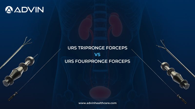
Triprong Forceps
Triprong Forceps feature three flexible prongs that open outward to grasp objects from multiple angles.
Key characteristics:
- Three-pronged design
- Moderate opening span
- Flexible structure
Clinical advantages:
- Good grip on irregular or round objects
- Balanced flexibility and control
- Easier maneuverability
They are commonly used in stone retrieval and general endoscopic grasping procedures.
Four-Prong Forceps
Four-Prong Forceps are designed with four prongs, offering wider coverage and enhanced grip stability.
Key characteristics:
- Four-point grasping design
- Wider opening capability
- Increased contact surface
Clinical advantages:
- Stronger and more stable grip
- Better hold on larger or slippery objects
- Reduced risk of slippage
Workflow-Based Usage: Balanced Grip vs Maximum Stability
Both forceps are used for retrieval but differ in gripping efficiency:
- Triprong Forceps → Balanced grip with flexibility
- Four-Prong Forceps → Maximum grip stability and coverage
This impacts:
- Holding strength
- Risk of slippage
- Object handling capability
Core Differences That Drive Clinical Choice
The key difference lies in number of contact points and grip strength:
- Triprong forceps offer flexibility and easier handling
- Four-prong forceps provide stronger grip and better stability
Quick Comparison Overview
| Aspect | Triprong Forceps | Four-Prong Forceps |
|---|---|---|
| Number of Prongs | 3 | 4 |
| Grip Type | Multi-point (moderate) | Multi-point (strong) |
| Stability | Good | Very high |
| Flexibility | High | Moderate |
| Contact Surface | Moderate | Larger |
| Slippage Risk | Moderate | Low |
| Object Size | Small to medium | Medium to large |
| Maneuverability | Easier | Slightly less |
Clinical Preference & Real-World Application
Triprong Forceps are commonly used in:
- Small to medium stone retrieval
- Irregular or mobile objects
- Procedures requiring flexibility
Their design ensures balanced grip with good maneuverability.
Four-Prong Forceps are preferred in:
- Larger stone retrieval
- Slippery or difficult-to-hold objects
- Situations requiring maximum grip
Practical Selection Guide
- Choose Triprong Forceps for flexibility and routine retrieval
- Choose Four-Prong Forceps for stronger grip and larger objects
- Use triprong for smaller, mobile stones
- Use four-prong for stable and firm holding
Conclusion: Flexibility vs Maximum Grip
Both instruments are essential in endoscopic retrieval:
- Triprong → Flexible, balanced, and easy to handle
- Four-Prong → Stable, strong, and high-grip performance
The ideal choice depends on:
- Object size and shape
- Required grip strength
- Procedural complexity
Why Advin Health Care is the Right Choice
Advin Health Care offers a wide range of retrieval forceps designed for precision and reliability.
- Triprong forceps provide versatile and efficient handling
- Four-prong forceps deliver strong grip and superior stability
With Advin, healthcare providers benefit from:
- High-quality materials
- Durable construction
- Consistent clinical performance
Get Connected:
+91-70717 27261 | urology@advinhealthcare.com | www.advinhealthcare.com