Thulium Laser Enucleation of Prostate Surgery Set – Complete Solution for Precision Laser Prostate Enucleation

Clinical Purpose of ThuLEP (Thulium Laser Enucleation of Prostate)

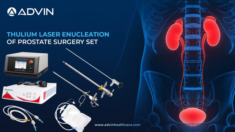
The ThuLEP Set is a comprehensive surgical solution designed for the treatment of benign prostatic hyperplasia (BPH) using advanced Thulium laser technology. It enables precise enucleation of enlarged prostate tissue with continuous laser emission, providing excellent cutting accuracy and superior hemostasis. This system integrates high-definition visualization, laser energy delivery, and efficient tissue removal to ensure safe, controlled, and effective prostate surgery.

Evolution of Thulium Laser Technology in Prostate Enucleation

Prostate surgery has progressed from open prostatectomy to minimally invasive techniques like TURP and laser-based procedures. While HoLEP introduced pulsed laser enucleation, Thulium laser technology offers continuous wave emission, allowing smoother and more controlled tissue dissection.

ThuLEP represents the next evolution in laser prostate surgery, providing precise incision, minimal bleeding, and improved visibility. Its continuous energy delivery allows surgeons to perform refined enucleation with enhanced control, making it an increasingly preferred technique in modern urology.

System Components and Functional Integration of ThuLEP Set

Equipment

  • Thulium Laser Machine
  • Advin HD Camera System
  • Medical Monitor 24″ / 27″
  • LED Light Source
  • Fiber Optic Cable

Instruments

  • TURP / Resectoscope
  • Laser Working Element
  • TURP Resectoscope Sheath 26 Fr
  • Urology Morcellator Set
  • Glass Ellik Bottle
  • Plastic Ellik Bottle

Disposables

  • Laser Fiber Optic
  • TUR Irrigation Set
  • Foley Balloon Catheter 3 Way 12–24 FG
  • Urine Collecting Bag

Drapes

  • TUR Drape
  • Lithotomy Drape
  • Under Buttock Drape

Clinical Applications of ThuLEP Procedure

  • Treatment of benign prostatic hyperplasia (BPH)
  • Enucleation of small to large prostate glands
  • Relief of urinary obstruction
  • Laser-based alternative to TURP and HoLEP
  • Precision prostate tissue removal with minimal bleeding

Instructions for Use of ThuLEP System

  • Set up the Thulium Laser Machine and connect the laser fiber optic for continuous laser energy delivery. 
  • Connect the Advin HD Camera System to the medical monitor, and attach the LED light source with fiber optic cable for clear visualization. 
  • Assemble the TURP resectoscope with 26 Fr sheath and insert the laser working element
  • Introduce the resectoscope through the urethra to access the prostate under direct vision. 
  • Start saline irrigation using the TUR irrigation set to maintain a clear surgical field. 
  • Use the thulium laser fiber to precisely enucleate prostate tissue layer by layer. 
  • Push the enucleated tissue into the bladder and remove it using the urology morcellator
  • Use the Ellik bottle if required for additional evacuation of tissue fragments. 
  • Insert a 3-way Foley catheter after the procedure for continuous irrigation and drainage. 
  • Connect to a urine collecting bag to monitor urine output post-surgery. 
  • Maintain sterility using TUR, lithotomy, and under buttock drapes throughout the procedure. 
  • After completion, clean and sterilize all reusable instruments properly.

Key Global Markets with High Adoption of ThuLEP Systems

  • Germany
  • Italy
  • Japan
  • India
  • South Korea

Clinical Advantages of ThuLEP Technology

  • Continuous wave laser for smooth and precise cutting
  • Excellent hemostasis with minimal bleeding
  • Enhanced visibility during surgery
  • Suitable for all prostate sizes
  • Reduced operative time in experienced hands
  • Faster recovery and shorter hospital stay

Advin Health Care ThuLEP Product Portfolio Overview

Advin Health Care offers a complete ThuLEP Set engineered for precision, control, and advanced laser prostate surgery. With high-performance Thulium laser systems, superior visualization, and reliable instrumentation, the set enables urologists to perform safe and efficient prostate enucleation while maintaining international quality standards.

Explore More Products>>

Get Connected:

+91-70717 27261 | urology@advinhealthcare.com | www.advinhealthcare.com

Suprapubic Cystostomy Surgery Set – Complete Urology Set for Suprapubic Catheterization Procedures

Clinical Purpose

The SPC Set is a specialized surgical solution designed for establishing suprapubic urinary drainage directly into the bladder through the abdominal wall. It is commonly used in cases where urethral catheterization is not possible or contraindicated. This set includes essential instruments, catheters, and disposables to ensure safe bladder access, efficient urine drainage, and improved patient comfort in both emergency and long-term urinary management.

Evolution of Suprapubic Catheterization

Suprapubic cystostomy has evolved from open surgical procedures to minimally invasive percutaneous techniques using trocar-based systems. Earlier approaches required larger incisions and longer recovery times.

Modern SPC techniques utilize guided trocar insertion, improved catheter materials (latex and silicone), and sterile procedural setups to enhance safety, reduce complications, and enable bedside or outpatient procedures. Today, SPC is widely used for long-term bladder drainage and in cases of urethral obstruction.

Set Components and Functional Integration

Instruments

  • SPC Trocar / Suprapubic Trocar – Metal

Disposables

  • Suprapubic Catheter Set (Latex / Silicone)
  • Foley Balloon Catheter 2 Way 12–24 FG
  • Urine Collecting Bag
  • Urine Leg Bag

Drapes

  • Cystoscopy Drape
  • Lithotomy Drape
  • Under Buttock Drape

Clinical Applications

  • Urinary retention when urethral catheterization fails
  • Urethral obstruction or injury
  • Long-term bladder drainage
  • Neurogenic bladder management
  • Post-operative urinary diversion

Instructions for Use (SPC – Suprapubic Cystostomy, Short & Smart Workflow)

  • Prepare the surgical area using cystoscopy, lithotomy, and under buttock drapes to maintain a sterile field. 
  • Identify the suprapubic entry site and ensure the bladder is adequately filled for safe access. 
  • Insert the SPC trocar (metal suprapubic trocar) through the lower abdominal wall into the bladder under controlled guidance. 
  • Once access is confirmed, introduce the suprapubic catheter (latex or silicone) through the trocar into the bladder. 
  • Remove the trocar carefully while keeping the catheter in place. 
  • Inflate the catheter balloon to secure it inside the bladder. 
  • Connect the catheter to a urine collecting bag or urine leg bag for continuous drainage. 
  • Use a Foley catheter additionally if required for urethral drainage support. 
  • Ensure proper fixation of the catheter to avoid displacement. 
  • After completion, dispose of single-use items and clean reusable instruments properly.

Countries with High Clinical Adoption

  • United States
  • Germany
  • India
  • United Kingdom
  • Middle East Countries

Key Clinical Advantages

  • Simple and minimally invasive procedure
  • Effective alternative to urethral catheterization
  • Suitable for long-term bladder management
  • Reduced risk of urethral trauma
  • Can be performed bedside or in OT
  • Improved patient comfort and mobility

Advin Health Care Product Overview

Advin Health Care offers a complete SPC Set designed for safety, reliability, and ease of use in suprapubic catheterization procedures. With high-quality trocars, durable catheter options, and essential disposables, the set enables healthcare professionals to perform efficient bladder drainage while maintaining international quality standards.

Explore More Products>>

Get Connected:

+91-70717 27261 | urology@advinhealthcare.com | www.advinhealthcare.com

Holmium Laser Enucleation of Prostate Surgery Set – Complete Solution for Advanced Laser Prostate Surgery

Clinical Purpose of HoLEP (Holmium Laser Enucleation of Prostate)

The HoLEP Set is a comprehensive surgical solution designed for the treatment of benign prostatic hyperplasia (BPH) using advanced Holmium laser technology. It enables precise enucleation of enlarged prostate tissue followed by efficient morcellation and removal. This system integrates high-power laser equipment, advanced visualization, and specialized instruments to ensure superior hemostasis, complete tissue removal, and improved patient outcomes in minimally invasive prostate surgery.

Evolution of Laser Technology in Prostate Enucleation (HoLEP Systems)

Prostate surgery has evolved from open prostatectomy to minimally invasive endoscopic techniques such as TURP. While TURP remains effective, it has limitations in managing large prostate glands.

The introduction of HoLEP marked a significant advancement, allowing complete anatomical enucleation of prostate tissue regardless of size. With the use of Holmium laser, surgeons achieve precise cutting, excellent hemostasis, and reduced bleeding. The addition of morcellation systems further enhances efficiency by enabling rapid removal of enucleated tissue. Today, HoLEP is considered a gold standard for large prostate management.

System Components and Functional Integration of HoLEP Set

Equipment

  • Holmium Laser Machine (30W / 60W / 100W / 120W / 160W)
  • Advin HD Camera System
  • USB Camera System
  • Endoscopy Video Recorder
  • Medical Monitor 24″ / 27″
  • LED Light Source
  • Fiber Optic Cable

Instruments

  • TURP / Resectoscope
  • Laser Working Element
  • TURP Resectoscope Sheath 26 Fr
  • Urology Morcellator Set
  • Glass Ellik Bottle
  • Plastic Ellik Bottle
  • Toomey Glass Syringe 50 ml / 100 ml

Disposables

  • Laser Fiber Optic
  • TUR Irrigation Set
  • Foley Balloon Catheter 3 Way Latex 12–22 FG
  • Foley Balloon Catheter 3 Way Silicone 16–24 FG
  • Urine Collecting Bag
  • Urometer

Drapes

  • TUR Drape
  • Lithotomy Drape
  • Under Buttock Drape
  • Universal Split Drape

Clinical Applications of HoLEP Procedure

  • Treatment of benign prostatic hyperplasia (BPH)
  • Management of large prostate glands
  • Complete prostate tissue enucleation
  • Relief of urinary obstruction
  • Alternative to TURP and open prostatectomy
Instructions for Use of HoLEP System
  • Set up the Holmium Laser Machine and connect the laser fiber optic for precise laser energy delivery. 
  • Connect the Advin HD Camera System or USB Camera System to the medical monitor, and attach the LED light source with fiber optic cable for clear visualization. 
  • Assemble the TURP resectoscope with 26 Fr sheath and insert the laser working element
  • Introduce the resectoscope through the urethra to access the prostate under direct vision. 
  • Start saline irrigation using the TUR irrigation set to maintain a clear surgical field. 
  • Use the holmium laser fiber to enucleate enlarged prostate tissue from the capsule. 
  • Push enucleated tissue into the bladder and use the urology morcellator to fragment and remove tissue. 
  • Use the Ellik bottle or Toomey syringe if required for additional evacuation of tissue fragments. 
  • Insert a 3-way Foley catheter after the procedure for continuous irrigation and drainage. 
  • Connect to a urine collecting bag or urometer to monitor urine output post-surgery. 
  • Maintain sterility using TUR, lithotomy, under buttock, and split drapes throughout the procedure. 
  • After completion, clean and sterilize all reusable instruments properly.

Key Global Markets with High Adoption of HoLEP Systems

  • United States
  • Germany
  • Japan
  • India
  • South Korea

Clinical Advantages of HoLEP Technology

  • Gold standard for large prostate treatment
  • Complete anatomical enucleation
  • Excellent hemostasis with minimal bleeding
  • Suitable for all prostate sizes
  • Reduced hospital stay and catheterization time
  • Lower reoperation rates compared to TURP

Advin Health Care HoLEP Product Portfolio Overview

Advin Health Care offers a complete HoLEP Set engineered for precision, efficiency, and advanced laser prostate surgery. With high-performance Holmium laser systems, superior visualization, and robust instrumentation, the set enables urologists to perform safe, effective, and modern prostate enucleation procedures while maintaining international quality standards.

Explore More Products>>

Get Connected:

+91-70717 27261 | urology@advinhealthcare.com | www.advinhealthcare.com

Optical Internal Urethrotomy Surgery Set – Comprehensive Solution for Urethral Stricture Management

Clinical Purpose of OIU (Optical Internal Urethrotomy)

The OIU Set is a specialized endoscopic solution designed for the treatment of urethral strictures through optical internal urethrotomy. It integrates high-definition visualization systems, precision urethrotomy instruments, and essential accessories to enable accurate incision of fibrotic urethral tissue, restoring normal urinary flow. This set ensures minimally invasive treatment, improved patient comfort, and effective clinical outcomes in urethral stricture management.

Evolution of Endoscopic Urethrotomy Technology

Urethral stricture management has evolved from open surgical reconstruction to minimally invasive endoscopic techniques. Traditional approaches involved extensive surgery with longer recovery times.

With the introduction of optical internal urethrotomy, surgeons can now directly visualize and incise strictures using endoscopic instruments. Modern advancements in optics, instrument design, and guidewire-assisted techniques have improved precision, reduced complications, and enhanced procedural success rates, making OIU a preferred first-line treatment in many cases.

System Components and Functional Integration of OIU Set

Equipment

  • Advin HD Camera System
  • LED Light Source

Instruments

  • Telescope
  • Optical Urethrotomy Sheath with Half Moon 21 Fr
  • OTIS Urethrotome
  • Cold Knife

Diposables

  • Guide Wire 0.028 / 0.032 / 0.035 / 0.038 – 150 cm

Clinical Applications of Optical Internal Urethrotomy

  • Treatment of urethral strictures
  • Restoration of urethral patency
  • Management of recurrent strictures
  • Minimally invasive alternative to open urethroplasty
  • Diagnostic evaluation combined with therapeutic incision
Instructions for Use of OIU System
  • Set up the Advin HD Camera System and connect it with the LED light source for clear endoscopic visualization. 
  • Assemble the telescope with optical urethrotomy sheath (21 Fr with half moon) and attach it to the camera system. 
  • Insert a guide wire through the urethra to safely identify and access the stricture. 
  • Advance the optical urethrotome (OTIS or sheath system) over the guide wire under direct vision. 
  • Use the urethrotome bridge to introduce the cold knife for precise incision of the stricture. 
  • Perform controlled incision at the stricture site to restore urethral patency. 
  • Ensure adequate opening and smooth passage through the urethra after incision. 
  • After completion, remove the instrument and place a catheter if required for healing support. 
  • Clean and sterilize all reusable instruments properly after the procedure. 

Clinical Advantages of OIU Technology

  • Minimally invasive urethral stricture treatment
  • Direct visualization for precise incision
  • Short procedure time
  • Reduced hospital stay
  • Cost-effective compared to open surgery
  • Quick patient recovery

Advin Health Care OIU Product Portfolio Overview

Advin Health Care offers a complete OIU Set designed for precision, safety, and efficiency in urethral stricture management. With high-quality optical systems and reliable urethrotomy instruments, the set enables urologists to perform accurate and effective procedures while maintaining international quality standards.

Explore More Products>>

Get Connected:

+91-70717 27261 | urology@advinhealthcare.com | www.advinhealthcare.com

PCNL Surgery Set with Holmium Laser System – Advanced Kidney Stone Removal Procedure Kit

Clinical Purpose of PCNL (Percutaneous Nephrolithotomy)

The PCNL Set with Laser is a comprehensive surgical solution designed for the treatment of large, hard, and complex kidney stones using a minimally invasive percutaneous approach combined with advanced laser lithotripsy. This system integrates high-definition visualization, precise renal access instruments, and high-power Holmium laser technology to enable effective stone fragmentation (dusting and fragmentation), safe extraction, and improved surgical outcomes.

Evolution of PCNL Technology with Laser Lithotripsy Systems

PCNL has evolved from open renal surgeries to a highly refined minimally invasive technique. Traditional PCNL relied primarily on pneumatic or ultrasonic lithotripters for stone fragmentation.

With the introduction of Holmium laser technology, PCNL procedures have achieved higher precision, better control over fragmentation (dusting vs fragmentation), and improved safety. Laser-assisted PCNL allows treatment of harder stones, reduces retropulsion, and enhances complete stone clearance. Combined with mini-PCNL advancements and improved imaging, it represents a modern, efficient approach to complex stone management.

System Components and Functional Integration of Laser PCNL Set

Equipment

  • Advin HD Camera System
  • LED Light Source
  • Fiber Optic Cable
  • Holmium Laser Machine (30W / 60W / 100W / 120W / 160W)
  • ADVIN C-ARM 3.5 KW / 5 KW

Instruments

  • R Wolf PCNL Nephroscope 12 Fr / 18 Fr / 24 Fr
  • PCNL Operative Sheath 12 / 20.8 / 24 Fr
  • Mini PCNL Sheath (All Sizes)
  • Alkine Cannula
  • Alkine Telescopic Dilator Set
  • Amplatz Sheath
  • Amplatz Sheath with Dilator
  • Amplatz Renal Dilator Set
  • Fascial Dilator Set
  • Nephrostomy Balloon Dilator
  • Inflation Device
  • PCNL Angular Basket
  • PCNL Forceps 5 / 6 / 7 / 8 / 9 Fr
  • PCNL Suction
  • PCNL Trocar
  • PCNL Tray

Dispsables

  • Laser Fiber Optic
  • Guide Wire 0.028 / 0.032 / 0.035 / 0.038 – 150 cm
  • PCN Catheter
  • PCN Catheter with Locking Mechanism
  • Malecot Nephrostomy Catheter
  • Double J Stent Set

Drapes

  • Urology Drape
  • Major Surgery Drape
  • Universal Split Drape

Clinical Applications of Laser-Based PCNL Procedure

  • Treatment of large and complex kidney stones
  • Management of hard calculi resistant to pneumatic lithotripsy
  • Staghorn calculi removal
  • Mini-PCNL procedures with laser fragmentation
  • Cases requiring precise stone dusting

Instructions for Use of Laser PCNL System

  • Set up the Advin HD Camera System, connect it to the medical monitor, and attach the LED light source with fiber optic cable for clear visualization. 
  • Position the Advin C-ARM to guide accurate kidney access under real-time imaging. 
  • Insert a guide wire into the kidney through percutaneous puncture to establish a safe access path. 
  • Dilate the tract using Alkine cannula, telescopic dilators, fascial dilators, or balloon dilator with inflation device
  • Place the Amplatz sheath (with or without dilator) to maintain a stable working channel. 
  • Introduce the PCNL nephroscope through the sheath to visualize renal stones. 
  • Connect the holmium laser machine and pass the laser fiber optic through the working channel for stone fragmentation. 
  • Fragment stones using the laser fiber under direct visualization. 
  • Remove stone fragments using PCNL forceps, angular basket, or suction system
  • Insert a PCN catheter or Malecot nephrostomy catheter for postoperative drainage. 
  • Place a Double J stent if required to support ureteral drainage and healing. 
  • Maintain sterility using urology, major surgery, and split drapes throughout the procedure. 
  • After completion, clean and sterilize all reusable instruments properly.

Key Global Markets with High Adoption of Laser PCNL Systems

  • United States
  • Germany
  •  Japan
  • India
  • South Korea

Advin Health Care Product Overview

Advin Health Care offers a complete PCNL Set with Laser designed for advanced, high-precision renal stone management. Combining robust nephroscopy instruments with powerful Holmium laser systems, the set enables urologists to perform efficient, safe, and modern PCNL procedures while maintaining international quality standards and optimized surgical performance.

Explore More Products>>

Get Connected:

+91-70717 27261 | urology@advinhealthcare.com | www.advinhealthcare.com

PCNL Surgery Set With Pneumatic Lithotripter – Complete Solution for Complex Kidney Stone Removal

Clinical Purpose of PCNL (Percutaneous Nephrolithotomy)

The PCNL Set with Pneumatic Lithotripter is a comprehensive surgical solution designed for the removal of large, complex, and staghorn kidney stones through a minimally invasive percutaneous approach. It integrates advanced imaging systems, nephroscopy instruments, dilation systems, and pneumatic lithotripsy technology to ensure efficient stone fragmentation, extraction, and renal access. This set enhances surgical precision, reduces operative time, and improves patient outcomes in complex urolithiasis cases.

Evolution of PCNL Technology with Pneumatic Lithotripsy Systems

PCNL has evolved from open kidney stone surgery to a highly advanced minimally invasive procedure. Earlier techniques required large incisions and longer recovery times.

With the introduction of nephroscopes, dilation systems (Amplatz, balloon dilators), and pneumatic lithotripters, PCNL has become the gold standard for large renal stones. Modern advancements, including mini-PCNL systems and improved imaging with C-arm guidance, have significantly reduced complications, improved accuracy, and enhanced patient recovery.

System Components and Functional Integration of PCNL Set

Equipment

  • Advin HD Camera System
  • LED Light Source
  • Fiber Optic Cable
  • Lithotripter – Advin Litho+
  • ADVIN C-ARM 3.5 KW / 5 KW

Instruments

  • R Wolf PCNL Nephroscope 12 Fr / 18 Fr / 24 Fr
  • PCNL Operative Sheath 12 / 20.8 / 24 Fr
  • Mini PCNL Sheath (All Sizes)
  • Alkine Cannula
  • Alkine Telescopic Dilator Set
  • Amplatz Sheath
  • Amplatz Sheath with Dilator
  • Amplatz Renal Dilator Set
  • Fascial Dilator Set
  • Nephrostomy Balloon Dilator
  • Inflation Device
  • PCNL Angular Basket
  • PCNL Forceps 5 / 6 / 7 / 8 / 9 Fr
  • PCNL Suction
  • PCNL Trocar
  • PCNL Tray

Disposables

  • Guide Wire 0.028 / 0.032 / 0.035 / 0.038 – 150 cm
  • PCN Catheter
  • PCN Catheter with Locking Mechanism
  • Malecot Nephrostomy Catheter
  • Double J Stent Set

Drapes

  • Urology Drape
  • Major Surgery Drape
  • Universal Split Drape

Clinical Applications of PCNL Procedure

  • Removal of large kidney stones (>2 cm)
  • Management of staghorn calculi
  • Treatment of complex or multiple renal stones
  • Cases where ESWL or RIRS is not effective
  • Percutaneous renal access and drainage

Instructions for Use of PCNL System

  • Set up the Advin HD Camera System, connect it to the medical monitor, and attach the LED light source with fiber optic cable for clear visualization.
  • Position the Advin C-ARM for real-time imaging guidance during renal access.
  • Insert a guide wire into the kidney through initial puncture to establish a safe access path.
  • Perform tract dilation using Alkine cannula, telescopic dilators, fascial dilators, or balloon dilator with inflation device.
  • Place the Amplatz sheath (with or without dilator) to maintain a stable working channel.
  • Introduce the PCNL nephroscope through the sheath to visualize the kidney stones.
  • Use the Advin Litho+ pneumatic lithotripter to fragment the stones under direct vision.
  • Remove stone fragments using PCNL forceps, angular basket, or suction system.
  • Insert a PCN catheter or Malecot nephrostomy catheter for drainage after the procedure.
  • Place a Double J stent if required to support ureteral drainage and healing.
  • Maintain sterility using urology, major surgery, and split drapes throughout the procedure.
  • After completion, clean and sterilize all reusable instruments properly.

Global Markets with High Adoption of PCNL Systems

  • United States
  • Germany
  • India
  • China
  • Brazil

Clinical Advantages of PCNL with Pneumatic Lithotripter

  • Gold standard for large and complex kidney stones
  • Highly effective stone clearance rate
  • Minimally invasive compared to open surgery
  • Advanced dilation options for safe renal access
  • Efficient fragmentation with pneumatic lithotripter
  • Reduced hospital stay and faster recovery

Advin Health Care PCNL Product Portfolio Overview

Advin Health Care offers a complete PCNL Set with Pneumatic Lithotripter designed for high-performance and precision in complex renal stone management. Equipped with advanced nephroscopy instruments, reliable dilation systems, and powerful lithotripsy technology, the set enables urologists to achieve efficient and safe outcomes while maintaining international quality standards.

Explore More Products>>

Get Connected:

+91-70717 27261 | urology@advinhealthcare.com | www.advinhealthcare.com

Retrograde Intrarenal Surgery Set – Complete Solution for Advanced Kidney Stone Management

Clinical Purpose of RIRS (Retrograde Intrarenal Surgery)

The RIRS Set is a comprehensive endoscopic solution designed for the diagnosis and treatment of kidney stones and intrarenal pathologies using flexible ureterorenoscopy. It integrates advanced laser systems, high-definition visualization, flexible scopes, and precision accessories to enable effective stone fragmentation and removal within the kidney. This set ensures minimally invasive treatment, enhanced patient safety, and superior clinical outcomes in modern urology.

Evolution of Flexible Endourology Technology in RIRS Systems

RIRS has evolved from limited-access ureteroscopic procedures to highly advanced intrarenal surgeries using flexible scopes and laser technology. Earlier techniques faced challenges in accessing all calyces and managing larger stones.

With the development of flexible ureterorenoscopes, improved deflection mechanisms, high-power Holmium and Thulium laser systems, and miniaturized accessories, RIRS has become a preferred modality for kidney stone management. These advancements allow precise stone dusting, better accessibility, reduced complications, and faster patient recovery.

System Components and Functional Integration of RIRS Set

Equipment

  • Flexible Ureterorenoscope (Reusable / Single Use)
  • Processor with Monitor for Disposable Scopes
  • Holmium Laser Machine (30W / 60W / 100W / 120W / 160W)
  • Thulium Laser Machine
  • Advin HD Camera System
  • Medical Monitor 24″ / 27″

Instruments

  • Ureteral Access Sheath
  • Laser Working Element

Disposables

  • Laser Fiber Optic
  • Guide Wire 0.028 / 0.032 / 0.035 / 0.038 – 150 cm
  • Hydrophilic Guide Wire
  • Stripped Guide Wire
  • Segura Basket 1.8 / 1.9 / 2.0 / 2.2 / 2.5 / 3 / 4 / 5 Fr × 120 cm
  • Helical Basket 2.5 / 3 / 4 / 5 Fr × 120 cm
  • N Compass Basket 3 / 4 Fr × 110 cm
  • Stone Cone 3 Fr × 115 cm
  • Double J Stent (Single / Multiloop / Silicone)

Drapes

  • Cystoscopy Drape
  • Lithotomy Drape
  • Under Buttock Drape
  • Universal Split Drape

Instructions for Use of RIRS System

  • Set up the flexible ureterorenoscope (reusable or single use) and connect it to the Advin HD Camera System or processor with monitor for clear visualization.
  • Prepare the Holmium or Thulium laser machine and connect the laser fiber optic through the working channel of the scope.
  • Insert a guide wire (PTFE / hydrophilic / Stripped) into the ureter to establish a safe access path.
  • Advance the ureteral access sheath over the guide wire to facilitate repeated entry and protect the ureter.
  • Introduce the flexible ureterorenoscope through the access sheath and navigate to the kidney to locate the stone.
  • Use the laser fiber to fragment the stone under direct visualization.
  • Retrieve stone fragments using Segura, Helical, or N Compass baskets as required.
  • Use a stone cone if needed to prevent stone migration during fragmentation.
  • Insert a Double J stent after the procedure to maintain ureteral drainage and healing.
  • Maintain sterility using cystoscopy, lithotomy, under buttock, and split drapes throughout the procedure.
  • After completion, clean and sterilize all reusable instruments properly.

Clinical Applications of RIRS Procedure

  • Treatment of kidney stones (renal calculi)
  • Management of lower, middle, and upper calyceal stones
  • Treatment of residual or recurrent stones
  • Intrarenal diagnostic evaluation
  • Management of ureteropelvic junction abnormalities
  • Stone dusting and fragmentation using laser

Key Global Markets with High Adoption of RIRS Systems

  • United States
  • Germany
  • Japan
  • India
  • South Korea

Clinical Advantages of RIRS Technology

  • Minimally invasive kidney stone treatment
  • Access to all intrarenal calyces
  • Advanced laser-based stone fragmentation (dusting technique)
  • Reduced hospital stay and faster recovery
  • Lower complication rates compared to open surgery
  • High success rate for complex renal stones

Advin Health Care RIRS Product Portfolio Overview

Advin Health Care offers a complete RIRS Set designed for precision, flexibility, and high-performance intrarenal procedures. With advanced laser compatibility, high-quality flexible ureterorenoscopes, and a full range of accessories, the system enables urologists to perform efficient and safe kidney stone management while adhering to international quality standards.

Explore More Products>>

Get Connected:

+91-70717 27261 | urology@advinhealthcare.com | www.advinhealthcare.com

Diagnostic Cystoscopy Set – Complete Solution for Accurate Lower Urinary Tract Evaluation

Clinical Purpose of Diagnostic Cystoscopy

The Diagnostic Cystoscopy Set is a comprehensive solution designed for the visual examination, diagnosis, and minor interventions within the lower urinary tract, including the urethra and bladder. It integrates advanced visualization systems, rigid and flexible cystoscopes, precision accessories, and essential disposables to ensure accurate diagnosis, patient safety, and efficient procedural workflow. This set is fundamental in urology for early detection and evaluation of urinary tract conditions.

Evolution of Cystoscopy Technology in Endoscopic Systems

Cystoscopy has evolved from basic rigid endoscopic systems to highly advanced flexible and HD imaging technologies. Traditional cystoscopy offered limited comfort and visualization, whereas modern systems provide enhanced optics, improved maneuverability, and better patient tolerance.

The introduction of flexible cystoscopes, high-definition camera systems, and minimally invasive accessories has significantly improved diagnostic accuracy, reduced patient discomfort, and enabled outpatient-based procedures. Today, cystoscopy is a routine and essential diagnostic tool in urology.

System Components and Functional Integration of Cystoscopy Set

Equipment

  • Advin HD Camera System
  • USB Camera System
  • LED Light Source
  • Medical Monitor 24″ / 27″

Instruments

  • Cystoscope 4 mm / 0°, 12°, 30° × 300 mm
  • Cystoscope 2.9 mm / 0°, 30° × 300 mm
  • Flexible Cystoscope (Reusable / Single Use)
  • Cystoscope Sheath with Obturator
  • Cystoscope Bridge (Single / Double)
  • Flexible Biopsy Forceps 4 / 5 / 6 / 7 Fr
  • Optical Biopsy Forceps
  • Cystoscopy Injection Needle

Disposables

  • Ureteric Catheter / Urethral Catheter (All Size Types)
  • Foley Balloon Catheter 2 Way 8–24 FG
  • Urine Collecting Bag

Drapes

  • Cystoscopy Drape
  • Lithotomy Drape
  • Under Buttock Drape

Clinical Applications of Diagnostic Cystoscopy

  • Diagnosis of hematuria (blood in urine)
  • Evaluation of bladder abnormalities (tumors, stones, inflammation)
  • Assessment of urethral strictures
  • Detection of urinary tract infections and lesions
  • Guidance for biopsy and minor endoscopic procedures
  • Follow-up and surveillance of urological conditions

Instructions for Use of Cystoscopy System

  • Set up the Advin HD Camera System or USB Camera System and connect it to the medical monitor for real-time visualization.
  • Attach the LED light source to the cystoscope using the fiber optic connection for proper illumination.
  • Assemble the cystoscope (rigid or flexible) with sheath and obturator for smooth and safe insertion.
  • Gently insert the cystoscope into the urethra and advance into the bladder for internal examination.
  • Use the cystoscope bridge to introduce instruments like biopsy forceps or injection needle when required.
  • Perform tissue sampling using flexible or optical biopsy forceps if abnormal areas are observed.
  • Use the cystoscopy injection needle for drug delivery or diagnostic purposes if needed.
  • Insert a ureteric or urethral catheter if required for drainage or access during the procedure.
  • After completion, place a Foley catheter if necessary and connect it to a urine collecting bag.
  • Maintain sterility using cystoscopy, lithotomy, and under buttock drapes throughout the procedure.
  • Clean and sterilize all reusable instruments after use.

Clinical Advantages of Diagnostic Cystoscopy

  • Minimally invasive diagnostic procedure
  • High-definition visualization for accurate diagnosis
  • Availability of rigid and flexible options
  • Enhanced patient comfort with flexible cystoscopy
  • Quick procedure with outpatient feasibility
  • Essential tool for early disease detection

Advin Health Care Cystoscopy Product Portfolio Overview

Advin Health Care offers a complete Diagnostic Cystoscopy Set designed for precision, clarity, and reliability in urological diagnostics. With advanced imaging systems and high-quality instrumentation, the set enables healthcare professionals to perform accurate evaluations and minor interventions while maintaining international quality and safety standards.

Explore More Products>>

Get Connected:

+91-70717 27261 | urology@advinhealthcare.com | www.advinhealthcare.com

Transurethral Resection of Bladder Tumor Set – Complete Solution for Safe and Precise Bladder Tumor Management

Clinical Purpose of TURBT (Bladder Tumor Resection)

The TURBT Set is a comprehensive surgical solution designed for the diagnosis, resection, and management of bladder tumors using transurethral endoscopic techniques. It integrates advanced electrosurgical systems, high-definition visualization, precision cutting instruments, and essential disposables to ensure accurate tumor removal, effective hemostasis, and optimal tissue sampling for histopathological evaluation. This set enhances procedural efficiency, safety, and clinical outcomes in urological oncology.

Evolution of TURBT Technology in Endoscopic Systems

Bladder tumor resection has evolved from basic cystoscopic interventions to advanced electrosurgical resection techniques. Early procedures relied on limited visualization and manual instruments, leading to incomplete resections and higher recurrence rates.

Modern TURBT systems now incorporate HD imaging, improved electrosurgical units, and a variety of specialized electrodes such as loop, roller, and Bugbee electrodes. The integration of bipolar energy and enhanced optics has significantly improved precision, reduced complications, and ensured better tumor staging and management.

System Components and Functional Integration of TURBT Set

Equipment

  • Electrosurgical Unit – Advin Electro+
  • Bipolar Saline TURP Generator – Advin BI TUR+
  • Advin HD Camera System
  • Medical Monitor 24″ / 27″
  • LED Light Source
  • Fiber Optic Cable

Instruments

  • Cystoscope 4 mm / 30° × 300 mm
  • Cystoscope Sheath with Obturator
  • Cystoscope Bridge (Single / Double)
  • TURP / Resectoscope
  • TURP Resectoscope Sheath 26 Fr
  • Cutting Loop
  • Ball Loop
  • Roller Loop
  • Collins Knife
  • Cold Knife
  • Bugbee Electrode 65 cm

Disposables

  • TUR Irrigation Set
  • Foley Balloon Catheter (2 Way / 3 Way) 12–24 FG
  • Urine Collecting Bag

Drapes

  • TUR Drape
  • Lithotomy Drape
  • Under Buttock Drape
  • Universal Split Drape

Clinical Applications of TURBT Procedure

  • Resection of bladder tumors (benign and malignant)
  • Diagnosis and staging of bladder cancer
  • Biopsy of suspicious bladder lesions
  • Control of bleeding in bladder tumors
  • Surveillance and follow-up procedures

Instructions for Use of TURBT System

  • Set up the Advin Electro+ Electrosurgical Unit or Advin BI TUR+ Bipolar Generator and connect it to the working element and electrodes for controlled cutting and coagulation.
  • Connect the Advin HD Camera System, Medical Monitor, LED Light Source, and Fiber Optic Cable to the cystoscope/resectoscope for clear visualization.
  • Begin with the cystoscope (4 mm, 30°) with sheath and obturator to inspect the bladder and identify the tumor.
  • Replace with the TURP resectoscope with 26 Fr sheath and attach the appropriate electrode (cutting loop, ball loop, roller loop, or Collins knife).
  • Connect the TUR irrigation set to maintain continuous fluid flow and clear visibility during the procedure.
  • Resect the bladder tumor using the cutting loop or Collins knife, and use ball or roller loop for coagulation.
  • Use the Bugbee electrode or cold knife when precise coagulation or incision is required.
  • Remove resected tumor tissue and ensure complete hemostasis under direct visualization.
  • Insert a 2-way or 3-way Foley catheter after the procedure for drainage and optional irrigation.
  • Connect to a urine collecting bag to monitor postoperative urine output.
  • After completion, place all reusable instruments in the sterilization tray and ensure proper cleaning.

Clinical Advantages of TURBT Technology

  • Complete endoscopic bladder tumor management solution
  • High precision tumor resection with multiple electrode options
  • Enhanced visualization with HD imaging systems
  • Effective hemostasis and reduced intraoperative bleeding
  • Supports accurate diagnosis and staging
  • Optimized workflow for urology operating rooms

Advin Health Care TURBT Product Portfolio Overview

Advin Health Care offers a complete TURBT Set engineered for precision, safety, and efficiency in bladder tumor management. Designed with advanced visualization systems and high-performance electrosurgical technology, the set enables urologists to perform accurate resections with confidence while ensuring compliance with international quality standards.

Explore More Products>>

Get Connected:

+91-70717 27261 | urology@advinhealthcare.com | www.advinhealthcare.com

TURP (Transurethral Resection of Prostate) – Monopolar Set: Complete Solution for Conventional Prostate Resection

Clinical Purpose of TURP (Transurethral Resection of Prostate) – Monopolar System

The TURP Monopolar Set is a comprehensive surgical solution designed for the effective management of benign prostatic hyperplasia (BPH) using monopolar electrosurgical energy. This system integrates electrosurgical units, visualization systems, precision resection instruments, and essential disposables to enable controlled prostate tissue removal, efficient hemostasis, and smooth procedural workflow. It remains a widely adopted and reliable technique in urological practice.

Evolution of TURP Technology in Monopolar Systems

Monopolar TURP is the traditional gold-standard technique for prostate resection and has been widely used for decades. It requires non-conductive irrigation fluids such as glycine to allow safe energy transmission. While effective, earlier systems carried risks like TUR syndrome and fluid imbalance.

With technological advancements, modern monopolar systems now offer improved energy control, enhanced visualization, and better safety mechanisms. Despite the rise of bipolar systems, monopolar TURP continues to be preferred in many clinical settings due to its proven efficacy, familiarity among surgeons, and cost-effectiveness.

System Components and Functional Integration of Monopolar TURP Set

Equipment

  • Bipolar Saline TURP Generator – Advin BI TUR+
  • Bipolar TURP + 400 W ESU (Advin BI TURP + ESU)
  • Electrosurgical Unit – Advin Electro+
  • ALL IN ONE Electrosurgical Unit (400 W + Safeseal + BITUR)
  • Advin HD Camera System
  • Medical Monitor 24″ / 27″
  • LED Light Source
  • Fiber Optic Cable

Instruments

  • TURP / Resectoscope
  • TURP Resectoscope Sheath 26 Fr
  • Monopolar Working Element (Single Active / Passive)
  • Monopolar TURP Cable / Monopolar L-Shape TURP Cable
  • Monopolar Loop Electrodes (Single Stem / Double Stem)
  • Glass Ellik Bottle / Plastic Ellik Bottle
  • Toomey Glass Syringe 50 ml / 100 ml
  • TURP Sterilization Tray

Disposables

  • TUR Irrigation Set (Endoscopic Irrigation Set)
  • Foley Balloon Catheter Latex 3 Way 12–22 FG
  • Foley Balloon Catheter Silicone 3 Way 16–24 FG
  • Urine Collecting Bag
  • Urometer

Drapes

  • TUR Drape
  • Lithotomy Drape
  • Under Buttock Drape
  • Universal Split Drape

Clinical Applications of Monopolar TURP Procedure

  • Treatment of Benign Prostatic Hyperplasia (BPH)
  • Resection of enlarged prostate tissue
  • Relief of urinary obstruction
  • Hemostasis in endoscopic urological procedures
  • Standard endoscopic prostate surgery

Instructions for Use of Monopolar TURP System

  • Set up the Advin Electro+ Electrosurgical Unit (ESU) and connect it with the monopolar cable and working element.
  • Connect the HD Camera System, Monitor, LED Light Source, and Fiber Optic Cable to the resectoscope for clear visualization.
  • Assemble the monopolar TURP resectoscope with sheath, then insert the working element with monopolar loop electrode.
  • Attach the irrigation set with non-conductive fluid (glycine) to maintain continuous flow and clear vision.
  • Perform prostate tissue resection using the monopolar loop electrode under direct visualization.
  • Remove tissue chips using the Ellik evacuator or Toomey syringe to keep the field clear.
  • Insert a 3-way Foley catheter after the procedure for drainage and bladder irrigation.
  • Connect to a urine bag or urometer to monitor urine output post-surgery.
  • After completion, place all reusable instruments in the sterilization tray for cleaning and sterilization.

Key Global Markets with High Adoption of Monopolar TURP Systems

  • India
  • United States
  • Germany
  • Brazil
  • Southeast Asian Countries

Clinical Advantages of Monopolar TURP Technology

  • Proven and widely accepted surgical technique
  • Cost-effective solution for healthcare facilities
  • High precision tissue resection
  • Reliable electrosurgical performance
  • Compatible with standard OT setups
  • Strong surgeon familiarity and ease of use

Advin Health Care Monopolar TURP Product Portfolio Overview

Advin Health Care offers a complete Monopolar TURP Set designed to deliver precision, reliability, and cost-effective performance in urological surgeries. Engineered with advanced electrosurgical technology and high-quality instrumentation, the system supports surgeons in achieving safe and efficient prostate resection outcomes while maintaining international quality standards.

Explore More Products>>

Get Connected:

+91-70717 27261 | urology@advinhealthcare.com | www.advinhealthcare.com