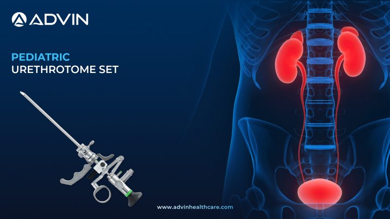
Introduction to the Optical Urethrotomy Set – Precision Instrument for Urethral Stricture Management
An Optical Urethrotomy Set is a surgical instrument used to treat urethral strictures by making a direct incision under endoscopic vision. It allows precise visualization and controlled cutting of the stricture to restore normal urine flow. This procedure is minimally invasive and effective for short-segment strictures.
Evolution of the Optical Urethrotomy Set – Advancements in Endoscopic Urethral Surgery
Optical internal urethrotomy was first introduced in the 1970s as a minimally invasive alternative to open urethroplasty. Early designs used cold knives under cystoscopic guidance. With advances in fiber optics and endoscopic technology, modern urethrotomy sets now offer superior visualization, integrated cold or laser knife options, and improved irrigation. The procedure remains one of the most widely performed initial treatments for male urethral strictures worldwide.
Understanding the Optical Urethrotomy Set and Its Key Components
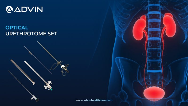
Advin Health Care offers a high-quality Optical Urethrotomy Set designed for accurate incision of urethral strictures under direct vision. The set includes a urethrotome sheath, working element, cold knife, and compatible telescope for endoscopic visualization. Manufactured from medical-grade stainless steel, it ensures durability, smooth operation, and optimal irrigation flow. Advin Health Care’s Optical Urethrotomy Set provides excellent precision, minimizing tissue trauma while restoring urethral patency effectively.
Clinical Applications – Effective Treatment of Urethral Strictures with Direct Vision
- Optical Internal Urethrotomy (OIU)
- Visual Internal Urethrotomy (VIU)
- Endoscopic Treatment of Urethral Stricture
- Urethral Dilation and Incision Procedures
- Post-Traumatic Urethral Obstruction Repair
How to Use the Optical Urethrotomy Set – Step-by-Step Surgical Guide for Urologists
- Assemble the urethrotome sheath, working element, and telescope
- Connect irrigation tubing for continuous visualization
- Introduce the instrument through the urethra under endoscopic guidance
- Identify the stricture site and insert the cold or laser knife
- Make a controlled incision at the 12 o’clock position of the stricture
- Ensure adequate dilation and confirm free urine flow
- Withdraw the instrument carefully and place a catheter if required
Clinical Benefits – Enhanced Visualization, Minimal Trauma & Faster Recovery
- Direct visualization ensures precise stricture incision
- Minimally invasive approach with short recovery time
- Reduces need for open urethroplasty in initial cases
- Compatible with both cold knife and laser techniques
- Allows complete irrigation for clear surgical view
- Durable and autoclavable components for repeated use
- Smooth assembly and ergonomic handling
- Effective treatment for anterior urethral strictures
Also Known As – Alternate Terms and Synonyms for Optical Urethrotomy Set
Optical Internal Urethrotome Set, Visual Internal Urethrotomy Set, Cold Knife Urethrotomy Set, VIU Instrument Set, OIU Surgery Kit, Endoscopic Urethral Stricture Set, Internal Urethral Cutting Instrument, Urethrotome Surgical Set, Optical Urethrotomy Equipment, Urethral Stricture Management Kit
Advin Optical Urethrotomy Set – Features, Components & Technical Specifications
- Advanced Endoscopic System for Internal Urethrotomy Procedures
- Advin Health Care is a trusted manufacturer of Optical Urethrotomy Set, providing precision-engineered, endoscopic-grade instruments designed for safe and effective urethral stricture management under direct vision.
- The Advin Health Care Optical Urethrotomy Set is designed for performing Direct Visual Internal Urethrotomy (DVIU) — a minimally invasive procedure used to treat urethral strictures under direct vision.
This precision-engineered set allows surgeons to visualize the urethral lumen clearly and incise strictures accurately, ensuring safe and effective treatment outcomes.
Advanced Features
- Excellent optical clarity for precise visualization
- Smooth cutting performance under direct vision
- Autoclavable & reusable stainless steel components
- Lightweight and ergonomic design for easy handling
- Compatible with standard 4mm 0° / 30° telescope
- International quality standard construction
The Optical Urethrotomy Set by Advin Health Care offers precision, durability, and reliability, making it the preferred choice for urologists performing urethral stricture management across the globe.
Get Connected:
+91-70717 27261 | urology@advinhealthcare.com | www.advinhealthcare.com